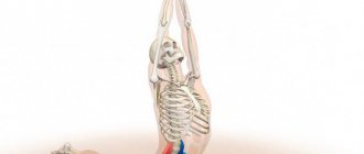
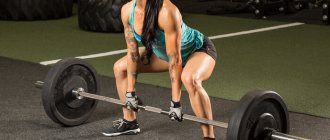
Muscles play a big role in the human body - they are the active part of our motor system. The passive part is formed by fascia, ligaments and bones. All skeletal muscles consist of muscle tissue: the torso, head and limbs. Their reduction occurs arbitrarily.
The muscles of the trunk and limbs, like the muscles of the head, are surrounded by fascia - connective tissue membranes. They cover areas of the body and get their name from them (fascia of the shoulder, chest, thigh, forearm, etc.).
About 40% of the total body weight of an adult is skeletal muscle. In children, they account for about 20-25% of body weight, and in older people - up to 25-30%. There are only about 600 different skeletal muscles in the human body. They are divided according to their location into the muscles of the neck, head, lower and upper extremities, as well as the torso (these include the muscles of the abdomen, chest and back). Let's take a closer look at the latter. We will describe the functions of the trunk muscles and give a name to each of them.
Intercostal external and internal muscles
The intercostal extrinsic muscles are located at all intercostal spaces from the costal cartilages to the spine. Their fibers go in the direction from top to bottom and forward. Since the lever of force (lever arm) is longer at the point of attachment of the muscle than at its origin, the muscles raise the ribs when they contract. Thus, the volume of the chest increases in the transverse and anteroposterior directions. These muscles are among the most important for inhalation. Their most dorsal bundles, which originate from the thoracic vertebrae (their transverse processes), stand out as the levator ribs muscles.
The internal intercostal spaces occupy about 2/3 of the anterior intercostal space. Their fibers go in the direction from bottom to top and forward. By contracting, they lower the ribs and thus facilitate exhalation, reducing the size of the human chest.
General information
Basic movement:
at degrees 5 and 4, rotation (rotation) and flexion of the body are performed simultaneously; at lower degrees, a pure rotation is performed. The range of motion of the thoracic region is 40°, the cervical region is 65°, of which 45° falls on the articulation between the atlas and axis. All degrees are tested in the supine position. Grade 2 can also be experienced in a sitting position. At degrees 5 and 4 we are talking about a combined movement of simultaneous rotation and flexion of the torso. Therefore, it is strictly taken into account that flexion and rotation occur simultaneously, while the torso, while bending (without extension or deflection in the lumbar region), is raised from the support.
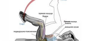
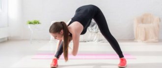
Muscles of the trunk involved in flexion with rotation
The movement throughout the entire length occurs at an equal speed, without jerking or swinging at the beginning. At degree 5, the arms maintain their original position throughout the entire movement; bending cannot be facilitated due to a shift in the center of gravity. During each test, posture is maintained using the arms. With degree 5, the hands are on the back of the head, with degree 4, the hands are along the body, with degree 3, they lie crosswise on the chest, with degrees 2, 1 and 0, they lie next to the body.
In grades 5 and 4 of the test, the lower limbs are strongly flexed to exclude involvement of the muscles that attach to the thighs. A squat like this is direct evidence of the involvement of the thigh muscles.
Pelvic fixation is certainly required to reduce hip involvement. During the entire movement we observe the state of the navel. When muscle tension is asymmetrical, it shifts towards the stronger muscle group.
When turning, for example, to the right, almost all the muscles of the body are activated, but to a greater extent the right internal oblique, left external oblique, right semispinalis, left multifidus, left rotator cuffs, left latissimus dorsi and right iliocostalis .
The rotation causes movement of the head in the cervical region (65°) and thoracic region (40°). In the lumbar region, due to the sagittal vertical arrangement of the joints of the spine, rotation is almost impossible. The amplitude of movement is limited by the position of the vertebral joints and the traction of the back muscles, and may also be due to the tension of the oblique abdominal muscles of the opposite side.
Table 1.4. Muscles of the trunk involved in flexion with rotation
Diaphragm
The thoracic obstruction (diaphragm) separates the abdominal cavity from the thoracic cavity. Even in the early period of embryonic development, this muscle is formed from the cervical myotomes. It moves back as the lungs and heart develop until it occupies a permanent position in a 3-month-old fetus. The diaphragm, according to the location of the anlage, is supplied with a nerve that arises from the cervical plexus. It is dome-shaped. The diaphragm consists of muscle fibers that begin around the circumference of the lower opening located in the chest. Then they pass into the tendon center, which occupies the top of the dome. The heart is located in the middle left part of this dome. In the thoraco-abdominal barrier there are special openings through which the esophagus, aorta, lymphatic duct, veins, and nerve trunks pass. It is the main respiratory muscle. When the diaphragm contracts, its dome lowers and the rib cage increases in vertical size. At the same time, the lungs are mechanically stretched and inhalation occurs.
Examples of antagonist muscles
The simplest examples of flexor and extensor muscles:
- The femoral biceps and quadriceps are the flexor and extensor muscles of the leg, or more precisely the hip. The biceps is located at the back, attached to the ischium at the top and bottom, turning into a tendon, adjacent to the femur in the area of the knee joint. And the quadriceps is an extensor, located on the front side of the thigh, attached by a tendon to the knee joint, and the upper part is attached to the pelvic bone.
- The biceps and triceps are the flexor and extensor muscles of the arm, located between the elbow and shoulder joints and attached to them by powerful tendons. They are the main muscles that form the shoulder and control the vast majority of flexion and extension movements of the arm.
You can often notice that if there is an overly active extensor muscle, then, as a result, the flexor muscle will be in a passive state, that is, not sufficiently developed, which creates inadequate body movements with a greater loss of energy than in harmoniously trained people (yogis are an example of this) .
Functions of the chest muscles
As you can see, the main function of the muscles listed above is to participate in the respiratory mechanism. Inhalation is caused by those that increase the volume of the chest. It occurs in different people either primarily through the diaphragm (the so-called abdominal type of breathing), or through the external intercostal muscles (thoracic type of breathing). These types can change, they are not strictly constant. The muscles that help reduce the volume of the chest are activated only with increased exhalation. For exhalation, the plastic properties possessed by the chest itself are usually sufficient.
Other chest muscles
The pectoralis major muscle originates from the edge of the sternum, the sternal part of the clavicle and the cartilages of the five or six upper ribs. It attaches to the humerus, the crest of its greater tubercle. Between it and the muscle tendon there is a synovial bursa. The muscle, contracting, pronates and adducts the shoulder, pulling it forward.
Below the major muscle is the pectoralis minor muscle. It starts from the second to fourth ribs, connects to the coracoid process and pulls the scapula downward and forward during contraction.
The serratus anterior muscle originates on the second to ninth ribs with nine teeth. It connects to the scapula (its medial edge and lower angle). The main part of its bundles is connected to the latter. When the muscle contracts, it pulls the scapula forward and its lower corner outward. Due to this, the scapula rotates around the sagittal axis, and the lateral angle of the bone rises. If the arm is abducted, rotating the scapula, the serratus anterior muscle raises the arm above the shoulder joint.
Posterior group - extensor muscles
Read:- I. Lateral muscle group of the hand
- I. Anterior group of shoulder muscles
- II. Back group
- III group.
- III group. Combined defects (6 patients).
- IV group
- A – rear surface; B – front surface.
- Algorithm for describing the manifestations of morbidity in population groups identified according to individual characteristics
- Art therapy in groups
- 1st group:
1. Triceps brachii muscle, i.e. triceps brachii,
located on the posterior surface of the humerus.
It starts with three heads. The long head, caput longum,
starts from the subarticular tubercle of the scapula;
lateral, caput laterale, -
from the posterior surface of the humerus and the lateral intermuscular septum;
medial, caput mediate, -
from the posterior surface of the humerus distal to the groove of the radial nerve and from the medial intermuscular septum. All the heads in the distal part are connected and attached to the olecranon process of the ulna.
Function: extends the forearm at the elbow joint. Innervation: radial nerve, Cv-CVII1.
2. Elbow muscle, t, anconeus,
triangular in shape, starting from the lateral epicondyle of the humerus; attaches to the posterior surface of the proximal end of the ulna. Function: extends the forearm at the elbow joint. Innervation: radial nerve, CVI-CVIII.
38. Brachial muscular canal, grooves of the shoulder and ulnar fossa, their purpose. The brachial canal (canalis humeromuscularis) is the space between the humerus and the heads of the triceps brachii muscle; the site of passage of the radial nerve, deep arteries and veins of the shoulder. The ulnar fossa (fossa cubitalis) is located in the anterior ulnar region, regio cubiti anterior. The ulnar fossa is limited by m. brachialis, medially – m. pronator teres, between them runs the medial (ulnar) groove, sulcus medialis (ulnaris). Laterally, the ulnar fossa is limited by m. brachioradialis. Between m. brachialis and m. brachioradialis there is a lateral (radial) groove, sulcus lateralis (radialis). The ulnar fossa contains superficial and deep vessels and nerves.
39. Muscles of the forearm, their functions, blood supply and innervation. Furrows of the forearm and their purpose. The forearm muscles are examined in a position of complete supination. According to their function, they are divided into two groups: anterior - flexors
and
pronators
and posterior -
extensors
and
supinators
.
Anterior group - flexors of the forearm and hand. 1. Brachioradialis muscle, t. brachioradialis,
starts from the lateral edge of the humerus and the lateral intermuscular septum; attaches to the lateral surface of the radius above the styloid process. Function: flexes the forearm and sets the radius in a mid-position between pronation and supination. Innervation: radial nerve, Cv-CVI.
2. Pronator teres, i.e. pronator teres,
starts from the medial epicondyle of the humerus and the medial intermuscular septum and ulnar tuberosity, goes down and laterally; attaches to the posterior edge of the radius above its middle. Function: pronates the forearm and participates in its flexion. Innervation: median nerve, CV|—CV1|.
3. Radial flexor carpi, t. flexor carpi radialis,
starts from the medial epicondyle of the humerus and fascia of the forearm; attaches to the base of the second metacarpal bone. Function: performs palmar flexion of the hand.
Innervation: median nerve, CV|—CV|1.
4. Long palmaris muscle, t. palmaris longus,
starts from the medial epicondyle and fascia of the forearm, forms a long tendon that passes into the palmar aponeurosis. Function: flexes the hand, strains the palmar aponeurosis. Innervation: median nerve, CV||—CVM|.
5. Flexor carpi ulnaris, t. flexor carpi ulnaris,
located medially. Starts from the medial epicondyle of the humerus, the fascia of the forearm, from the olecranon process and the posterior curve of the ulna; attaches to the pisiform bone. Function: bends and adducts the hand. Innervation: ulnar nerve, CVII—CVI||.
The listed 5 muscles make up the superficial layer of the forearm flexors. Deeper lie 4 muscles, forming a deep layer.
1. Superficial flexor of the fingers, i.e. flexor digitorum superficialis,
starts from the medial epicondyle of the humerus, the coronoid process of the ulna and the anterior surface of the radius. At the distal end, the muscle forms 4 tendons that pass through the carpal tunnel to the hand. The tendons are attached to the lateral surface of the middle phalanges of the II-V fingers. Function: bends the middle phalanges of the II-V fingers and the hand. Innervation: median nerve, CVM-CVI1|.
2. Long flexor of the thumb, hand, t. flexor pollicis longus,
begins on the anterior surface of the radius, interosseous membrane; attaches to the base of the distal phalanx of the thumb.
Function: flexes the distal phalanx of the thumb. Innervation: median nerve, Cvl-CV||.
3. Deep flexor of the fingers, t. flexor digitorum profundus,
starts from the anterior surface of the ulna and the interosseous membrane of the forearm. At the distal end of the forearm it forms 4 tendons, which pass in the carpal canal along with the tendons of the superficial flexor of the fingers and are attached to the bases of the distal phalanges of the II-V fingers. Function: flexes the distal phalanges of the fingers and hand. Innervation: median and ulnar nerves, CV||—Thr
4. Square pronator, i.e. pronator quadratus,
located in the distal forearm, lies under the deep flexor of the digitorum. Starts from the anterior and partially posterior surfaces of the ulna; attaches to the lateral and anterior surfaces of the radius. Function: rotates the radius inward. Innervation: median nerve, CVI—Thr
Posterior group - extensors of the forearm and hand. Arranged in two layers - superficial
and
deep.
Surface layer. 1. Extensor carpi radialis longus, i.e. extensor carpi radialis longus,
starts from the lateral edge and from the lateral epicondyle of the humerus; attaches to the base of the second metacarpal bone.
Function: flexes the forearm, extends and abducts (together with the so-called flexor carpi radialis)
brush.
Innervation: radial nerve, CV|—CVM.
2. Extensor carpi radialis brevis, i.e. extensor carpi radialis brevis,
starts from the lateral epicondyle of the humerus; attaches to the base of the third metacarpal bone. Function: extends the hand.
Innervation: radial nerve, CV|-CVM1.
3. Extensor digitorum, i.e. extensor digitorum,
starts from the lateral epicondyle of the humerus, in the distal section it is divided into 4 tendons, which pass under the extensor retinaculum,
retinaculum extensorum,
go to the dorsum of the II-V fingers and are attached to the distal and middle phalanges.
Function: extends fingers II-V. Innervation: radial nerve, CV1—CVI1|. t
4.
The extensor of the little finger, m.extensor digiti minimi,
is separated from the extensor of the fingers and is attached to the base of the distal phalanx of the fifth finger. 1 Function: extends the V finger.
Innervation: radial nerve, CV|—CV
Extensor carpi ulnaris, i.e. extensor carpi ulnaris, starts from the lateral epicondyle of the humerus and the posterior edge of the ulna, attaches to the base of the fifth metacarpal bone. Function: extends and adducts (together with the m. flexor carpi ulnaris) the hand. Innervation: radial nerve, CV||—Cvlir Deep layer. 1. Arch support, i.e. supinator, starts from the lateral epicondyle of the humerus and from the crest of the ulnar supinator. Attaches to the radius. Function: rotates the radius outward. Innervation: radial nerve, Cv-CV|. 2. Long muscle, abductor pollicis, i.e. abductor pollicis longus, starts from the distal parts of the bones of the forearm and the intermuscular membrane; attaches to the base of the first metacarpal bone. Function: abducts the thumb. Innervation: radial nerve, CVI-CV||. 3. Short extensor of the thumb, i.e. extensor pollicis brevis, starts from the posterior surface of the radius and the interosseous membrane; attaches to the main phalanx of the thumb. Function: extends and abducts the thumb. Innervation: radial nerve, CV|—CV1I. 4. Extensor pollicis longus, i.e. extensor pollicis longus, starts from the posterior surface of the ulna, interosseous membrane; attaches to the posterior surface of the distal phalanx of the thumb. Function: extends the thumb. Innervation: radial nerve, CV1—CV1|. 5. Extensor of the index finger, i.e. extensor indicis, starts from the posterior surface of the ulna, near the head; attaches to the extensor tendon of the digitorum, which goes to the index finger. Function: extends the 11th finger. Innervation: radial nerve, CV||—CV11|. 40. Muscles of the hand, their functions, blood supply and innervation. Osteofibrous canals and synovial sheaths of the hand. There are short muscles on the hand, which form three groups on the palmar surface: lateral, medial and middle. Lateral group. It consists of 4 muscles: the short muscle, abductor pollicis, i.e. abductor pollicis brevis; flexor pollicis brevis, i.e. flexor pollicis brevis; muscle that opposes the thumb to the hand, i.e. opponens pollicis; adductor pollicis muscle, i.e. adductor pollicis. All muscles originate from the carpal bones and the flexor retinaculum; attached to the base of the proximal first phalanx. Function: corresponds to the names of the muscles. Innervation: short flexor and adductor muscle - ulnar nerve, CV1I-Th; short, abducens and opposites - median nerve, CVI-Cvn. Medial group. The muscles of this group are less developed than the lateral group. It consists of 4 muscles: the muscle that abducts the little finger, i.e. abductor digiti minimi; short flexor of the little finger, t. flexor digiti minimi brevis; muscle opposite the little finger, i.e. opponens digiti minimi. They start from the flexor retinaculum and carpal bones; attached to the proximal phalanx of the little finger and fifth metacarpal bone. This group also includes the palmaris brevis muscle, i.e. palmaris brevis. Function: corresponds to the names of the muscles. Innervation: ulnar nerve, CV||—Thr Middle group. The lumbrical muscles belong to this group, mm. lumbricales; palmar and dorsal interosseous muscles, mm. interossei palmares et dorsales. Function: vermiforms bend the proximal phalanges of the II-V fingers; palmar interosseous brings the fingers together, flexes the first phalanges and extends the second and third phalanges; the dorsal ones spread the fingers, bend I-IV and push apart the II-III phalanges. Innervation: median, ulnar nerves, CVII1—Th. 41. Pelvic muscles, their functions, blood supply and innervation. Obturator canal, supra- and infrapiriform foramina, their walls and contents. The muscles of the lower limb girdle - the pelvis - surround the hip joint. They start from the sacrum, pelvic bones and spine; are attached to the proximal end of the femur. Topographically, they are divided into two groups: internal and external pelvic muscles. INTERNAL MUSCLES OF THE PELVIS 1. Iliopsoas muscle, i.e. iliopsoas, consists of two muscles: the iliacus, i.e. iliacus, starting in the iliac fossa, and the lumbar major, i.e. psoas major, originating from the bodies of the XII thoracic and I-II lumbar vertebrae and the transverse processes of the latter. Both muscles join together, pass under the inguinal ligament in the muscle lacuna and are attached to the lesser trochanter of the femur. Function: flexes the thigh and rotates it outward. Innervation: lumbar plexus, L,—Llv. 2. Psoas minor muscle, i.e. psoas minor, unstable, starts from the bodies of the XII thoracic and I lumbar vertebrae; attaches to the fascia iliaca. Function: stretches the fascia iliaca. Innervation: lumbar plexus, L,—L|r 3. Piriformis muscle, i.e. piriformis, starts from the pelvic surface of the sacrum, passes through the greater sciatic foramen; attaches to the greater trochanter of the femur. Function: rotates the thigh outward. Innervation: sacral plexus, S,—S,,. 4. Obturator internus muscle, i.e. obturatorius internus, starts from the inner surface of the obturator membrane and the inner surface of the pelvic bone around the obturator foramen; attaches to the trochanteric fossa. Function: rotates the thigh outward. Innervation: sacral plexus, L,—S|r 5. Superior and inferior gemellus muscles, mm. gemelli superior et inferior, start from the ischial spine (upper) and the ischial tuberosity (lower); are attached in the trochanteric fossa. Function: rotate the thigh outward. Innervation: sacral plexus, L|V—S|r external pelvic muscles 1. Gluteus maximus, i.e. glutens maximus, starts from the outer surface of the ilium behind the upper gluteal line, from the dorsal surface of the sacrum and coccyx; attaches to the gluteal tuberosity of the femur and the iliotibial tract of the lata fascia of the thigh. Function: extends the hip, rotates outward, fixes the pelvis. Innervation: inferior gluteal nerve, LM—L|v. 2. Gluteus medius and minimus, mm. glutei medius et minimus, located under the gluteus maximus muscle. Start from the outer surface of the ilium, between the anterior and posterior gluteal lines; are attached to the greater trochanter of the femur. Function: the thigh is abducted, the anterior bundles are rotated inward, the posterior bundles are rotated outward. With fixed lower limbs, tilt the pelvis to the side. Innervation: superior gluteal nerve, L|V-S. 3. Tensor fascia lata, i.e. tensor fasciae latae, located on the outer surface of the thigh. It starts from the iliac crest, from the superior external iliac spine, goes down and passes into the iliotibial tract, tractus iliotibialis, which attaches to the lateral condyle of the tibia. Function: strains the fascia lata, flexes the hip and rotates it inward. Innervation: superior gluteal nerve, L1V-S. 4. Quadratus femoris muscle, i.e. quadratus femoris, starts from the ischial tuberosity; attaches to the intertrochanteric ridge. Function: rotates the thigh outward. Innervation: cross plexus, LIV-S. 5. The external obturator muscle, m.obturatorius externus, starts from the outer surface of the pelvic bone, from the obturator membrane; attaches to the trochanteric fossa. Function: rotates the thigh outward. Innervation: obturator nerve, L...-LV 42. Thigh muscles, their functions, blood supply and innervation. There are three groups of muscles on the thigh: anterior - extensor muscles, posterior - flexor muscles and medial - adductor muscles. The anterior group is the extensor muscles. 1. Quadriceps femoris muscle, i.e. quadriceps femoris, located on the front surface of the thigh and consists of 4 heads - muscles. Rectus femoris muscle, i.e. rectus femons, lies superficially on the vastus intermedius; starts from the inferior anterior iliac spine; vastus medialis muscle, i.e. vastus medialis, originates from the medial lip of the linea aspera, and the vastus lateralis muscle, i.e. vastus lateralis, - from the lateral lip of the linea aspera; vastus intermedius muscle, i.e. vastus intermedius, starts from the anterior surface of the thigh. In the distal section, all heads of the quadriceps femoris muscle pass into the common tendon covering the patella and are attached to the tibial tuberosity. Function: extends the lower leg at the knee joint, m. The rectus femoris flexes the thigh. Innervation: femoral nerve. 2. Sartorial muscle, i.e. sartorius, starts from the superior anterior iliac spine; attaches to the medial surface of the tibial tuberosity. Function: flexes the thigh and lower leg, rotates the limb bent at the knee joint inward. Innervation: femoral nerve. Posterior group - extensor muscles. 1. Biceps femoris muscle, m. biceps femoris, has two heads, occupies a lateral position. The short head, caput breve, begins from the distal part of the lateral lip of the linea aspera, the long head, caput longum, from the ischial tuberosity. Both heads form a common belly, which is attached to the head of the fibula. Function: with a fixed pelvis, flexes the lower leg at the knee joint and extends the thigh. Innervation: sciatic nerve, L1V-S. 2. Semitendinosus muscle, i.e. semitendinosus, located on the medial surface of the thigh. It starts from the ischial tuberosity, in the middle part it is interrupted by a tendon jumper; attaches to the tibial tuberosity. Function: with a fixed pelvis, flexes the lower leg and extends the thigh. Innervation: sciatic nerve. 3. Semimembranosus muscle, i.e. semimetnbranosus, starts from the ischial tuberosity with a lamellar tendon, which is half the length of the muscle; attaches to the medial condyle of the tibia. Function: with a fixed pelvis, extends the thigh, bends and turns the lower leg inward. Innervation: sciatic nerve. Medial group - adductor muscles. 1. Pectineus muscle, i.e. pectineus. 2. Long adductor muscle, i.e. adductor longus. Starts from the superior branch of the pubic bone. 3. Thin muscle, i.e. gracilis. 4. Short adductor muscle, i.e. adductor brevis. Originates from the lower branch of the pubic bone. 5. Adductor magnus muscle, i.e. adductor magnus, starts from the ischial tuberosity and the anterior surface of the lower branches of the ischium and pubis. All of these muscles are attached to the medial lip, and the gracilis muscle is attached to the tibial tuberosity. Function: adduct and flex the hip; the thin muscle flexes the lower leg and rotates it inward. Innervation: obturator nerve. 43. Muscular and vascular lacunae, femoral and adductor canals, their walls, openings and contents. In the subfascial layer of the anterior thigh under the inguinal ligament there are muscular and vascular lacunae, lacuna musculorum and lacuna vasorum. The muscular lacuna corresponds to the outer 2/3 of the inguinal ligament and is separated from the vascular lacuna by the tendinous iliopectineal arch, arcus iliopectineus, running from the inguinal ligament to the iliopubic eminence, eminentia iliopubica. Muscle lacuna. Walls of the muscle lacuna The walls of the muscle lacuna are: anteriorly - the inguinal ligament, posteriorly - the crest of the pubic bone, medially - the arcus iliopectineus. Through the muscle lacuna, m. emerge onto the anterior surface of the thigh. iliopsoas and femoral nerve, n. femoralis (branch of the lumbar plexus). The walls of the vascular lacuna are: in front - the inguinal ligament, behind - the crest of the pubic bone, laterally - the tendinous arch, medially - the lacunar, or gimbernate, ligament, lig. lacunare. The femoral artery and vein pass through the lacuna vasorum (the vein is located medially, and the artery is located laterally), as well as the femoral branch of the genitofemoral nerve. The femoral artery can be pressed against the bone here to temporarily stop bleeding if it is damaged. Inward from the vessels (v. femoralis) is the femoral ring, anulus femoralis, which is the deep opening of the femoral canal. The adductor canal is a continuation of the anterior groove of the femur at the border of the middle and lower third. The skin in the medial part of the adductor canal region is thin and mobile; outward it thickens and is firmly fixed to the underlying tissues. In a well-developed layer of subcutaneous tissue there is (in the form of one or two trunks) the large saphenous vein of the leg, v. saphena magna. Rr. cutanei anteriores (n. femoralis) penetrate through the fascia lata along the inner edge of m. sartorius and spread in the skin of the anterior thigh up to the patella. The cutaneous branch of the obturator nerve penetrates the fascia lata in the middle of the medial thigh and reaches the patella. The adductor canal (canalis adductorius) is located under the fascia lata and is covered in front by m. sartorius. The medial wall of the adductor canal is m. adductor magnus, lateral wall of the adductor canal - m. vastus medialis. The anterior wall of the adductor canal is formed by a broad adductor intermuscular septum, septum intermusculare vastoadductoria, stretched from the adductor magnus muscle to the m. vastus medialis. There are three openings in the adductor canal. Through the upper opening from the sulcus femoralis anterior, the femoral vessels and n. pass into the canal. saphenus. The lower hole is a gap between the bundles of the adductor magnus muscle or between its tendon and the femur; through it the femoral vessels pass into the popliteal fossa. The anterior opening in the septum intermusculare vastoadductoria is the place of exit from the canal (into the tissue under the m. sartorius) of the descending knee artery and vein, a. et v. descendens genus and n. saphenus. Vessels and p. saphenus can exit the canal separately; in these cases there will be multiple front holes. The length of the adductor canal (canalis adductorius) is 5-6 cm, its middle is 15-20 cm from the tuberculum adductorium femoris on the medial epicondyle of the femur. In the proximal direction, the adductor canal communicates with the space of the femoral triangle, distally - with the popliteal fossa, along a et v. descendens genus and p. saphenus - with subcutaneous tissue on the medial surface of the knee joint and lower leg. According to these connections, purulent processes may spread in this area. The fascial sheath of the femoral vessels is firmly fused with the upper edge of the septum intermusculare vastoadductoria, and below the vessels deviate from this plate by 1.0-1.5 cm, with the femoral artery lying anteriorly and medially, and the vein posteriorly and laterally. A. descendens genus (single or double) reaches the arterial network of the knee joint, sometimes forming a direct anastomosis with the anterior recurrent branch of the tibial artery, a. recurrens tibialis anterior. N. saphenus in the subcutaneous cell of the leg joins v. saphena magna and reaches the middle of the inner edge of the foot. The femoral canal is located between the superficial and deep layers of the fascia lata. The femoral canal has two openings - deep and superficial, and three walls. The deep opening of the femoral canal is projected onto the inner third of the inguinal ligament. The superficial opening of the femoral canal, or subcutaneous fissure, hiatus saphenus, is projected 1-2 cm downward from this part of the inguinal ligament. The hernia emerging from the abdominal cavity penetrates the canal through a deep hole - the femoral ring, anulus femoralis. It is located in the very medial part of the vascular lacuna and has four edges. In front, the femoral ring is limited by the inguinal ligament, and behind by the pectineal ligament, lig. pectineale, located on the crest of the pubic bone (pecten ossis pubis), medially - lacunar ligament, lig. lacunare, located in the angle between the inguinal ligament and the crest of the pubic bone. On the lateral side it is limited by the femoral vein. The femoral ring faces the pelvic cavity and is covered on the inner surface of the abdominal wall by the transverse fascia, which here looks like a thin plate, septum femorale. Within the ring there is a deep inguinal lymph node. The superficial ring of the femoral canal (orifice) is the subcutaneous fissure, hiatus saphenus, a defect in the superficial layer of the fascia lata. The opening is closed by the cribriform fascia, fascia cribrosa. The walls of the femoral canal are a triangular pyramid. The anterior wall of the femoral canal is formed by a superficial layer of fascia lata between the inguinal ligament and the upper horn of the subcutaneous fissure - cornu superius. The lateral wall of the femoral canal is the medial semicircle of the femoral vein. The posterior wall of the femoral canal is a deep layer of fascia lata, which is also called fascia iliopectinea. There is no medial wall of the femoral canal, since the superficial and deep layers of fascia of the long adductor muscle grow together. The length of the femoral canal (the distance from the inguinal ligament to the superior horn of the hiatus saphenus) ranges from 1 to 3 cm. 44. Muscles of the lower leg, their functions, blood supply and innervation. The ankle-popliteal canal, its walls, openings, contents. The lower leg muscles are divided into three groups: anterior, posterior and lateral. Front group. 1. Tibialis anterior muscle, i.e. tibialis anterior. Starts from the lateral condyle and the lateral surface of the tibia and the interosseous membrane of the tibia; attaches to the medial cuneiform bone and the base of the first metatarsal bone. Function: extends and supinates the foot. Innervation: deep peroneal nerve. 2. Extensor digitorum longus, i.e. extensor digitorum longus, lies lateral to the previous muscle. It starts from the lateral condyle of the tibia, from the head and anterior edge of the fibula and the interosseous membrane of the tibia. The muscle is divided into 5 tendons, four of which are attached to the distal phalanges of the II-V fingers, the fifth - to the V metatarsal bone. • Function: extends fingers and foot. Innervation: deep peroneal nerve. 3. Extensor pollicis longus, i.e. extensor hallucis longus, starts from the lower part of the medial surface of the fibula and the interosseous membrane; attaches to the distal phalanx of the first finger. Function: extends the big toe, extends and supinates the foot. Innervation: deep peroneal nerve. Back group. 1. Triceps surae muscle, i.e. triceps surae, forms the surface layer. Consists of the gastrocnemius muscle, i.e. Gastrocnemius, which begins with two heads from the medial and lateral condyles of the femur, and the kam-back muscle, t. soleus, extending from the proximal departments of the bones of the lower leg and the tendon arc. Both muscles, connecting, form a powerful heel [Achilles] tendon, tendo calcaneus, attached to the heel of the hill. Function: bends the foot and lower leg. Innervation: tibia nerve. 2. The subsidized muscle, t. Plantaris, starts from the popliteal surface of the femur, passes into a long tendon, which attaches to the heel bone. Function: pulls the capsule of the knee joint posteriorly. Innervation: tibia nerve. 3. Podged muscle, t. Popliteus, forms along with the subsequent deep layer. Originates from the lateral label of the femur; It is attached to the proximal epiphysis of the tibia. Function: bends the lower leg and rotates it inside. Innervation: tibia nerve. 4. A long flexor of the thumb of the foot, t. Flexor Hallucis Longus, begins on the rear surface of the fibula and from the inter -cell membrane, passes behind the medial ankle; It is attached to the distal phalanx of the first finger. Function: I begs I finger. Innervation: tibia nerve. 5. A long flexion of the fingers, t. Flexor Digitorum Longus starts from the posterior surface of the tibia, passes behind the medial ankle and is divided into 4 tendons on the sole, which are attached to the distal phalanges of the II-V of the fingers. Function: bends the distal phalanges of the II - V of the fingers, and also bends the foot and skimpers. Innervation: tibia nerve. 6. The back of the tibial muscle, t. Tibialis Posterior, lies under the previous muscles. It begins from the proximal epiphesis of the bones of the lower leg, the inter -cell membrane, envelops the medial ankle, passes to the foot; It is attached to the tuberosity of the scaphoid, three wedge -shaped bones and bases of the II -V Plus Bones. Function: Bends and skimpers the foot. Innervation: tibia nerve. Lateral group. 1. The long unimportant muscle, t. Fibularis longus, begins from the head of the fibula; It is attached to the medial wedge -shaped and I plus bones. Function: raises the lateral edge of the foot, at the same time lowers the medial edge, bends the foot. 2. A short fibula, t. Fibularis brevis, starts from the fibula; It is attached to the bugrostism V of the plus bone. Function: bends the foot, raises its lateral edge. Innervation: both muscles are innervated by a superficial hearse nerve. 45. The muscles of the foot, their functions, blood supply and innervation. Subanary intermuscular furrows. Synovial vagina of the foot. Distinguish between the muscles of the rear and the muscles of the sole of the foot. On the rear, two muscles are stopped: a short extensor of the fingers, t. Extensor Digitorum Brevis, and a short extensor of the thumb of the foot, t. Extensor Hallucis Brevis. Both muscles start from the heel, attach to the phalanges of the I - V fingers. Function: Finger extend. Innervation: deep fiber nerve. Three groups are divided into podsmys. The medial group consists of a muscle diverting the thumb of the foot, t. abdUctor Hallucis; short flexor of the thumb of the foot, t. Flexor Hallucis Brevis; The muscles leading the thumb of the foot, t. AddUctor Hallucis. The lateral group includes a short flexor of the little finger of the foot, t. Flexor digiti minimi brevis; The muscle diverting the little finger of the foot, t. ABDUCTOR Digiti Minimi. The middle group includes a short flexion of the fingers, t. Flexor Digitorum Brevis; The square muscle of the sole, t. Quadratus Plantae; Cherve -shaped muscles, mm. lumbricales; Social inter -ocatual muscles, mm. Interossei Plantares, and back intercosal muscles, mm. Interossei Dorsales. Function: The worm -shaped muscles bend the phalanges of the fingers, the inter -regional rear - push the extends, and the inter -cellular plantar - shift the fingers. Innervation: medial and lateral plantar nerves.
Date added: 2014-12-11 | Views: 560 | Copyright infringement
| | | | | | 7 | | | | | | | | | | |
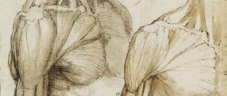
Rectus and pyramidalis muscles
The rectus abdominis muscle begins from the cartilages of the fifth to seventh ribs, as well as the xiphoid process. It is attached to the pubic symphysis outward from it. This muscle is intercepted transversely using 3 or 4 tendon bridges. The rectus muscle is located in a fibrous sheath formed by the aponeuroses of the oblique muscles.
The next one, the pyramidal muscle, is small, often completely absent. It is a vestige of the bursa muscle found in mammals. It begins near the pubic symphysis. This muscle, tapering upward, attaches to the linea alba, tightening it when contracted.
Errors and instructions
1. The movement occurs incorrectly if the patient tries to quickly make a movement with a large swing (jerk) and if he lifts the entire body “like a board,” which causes lordosis in the lumbar region.
2. This also occurs if the required maximum flexion of the lower limbs is not maintained.
3. With weak oblique abdominal muscles, the patient first tries to bend himself and only at the end of the movement rotates the torso through a sharp jerk.
4. If the quadratus lumborum muscle is highly developed (its activity predominates), the patient tries to simultaneously do unilateral flexion.
5. If the elbow joints are not maximally separated from each other during movement corresponding to degree 5, then they cannot support flexion or rotation.
External and internal oblique muscles
The external oblique originates from the lower ribs in eight bundles. Its fibers go from top to bottom and forward. This muscle attaches to the ilium (its crest). In front it passes into the aponeurosis. The fibers of the latter participate in the formation of the rectus sheath. They intertwine along the midline with the fibers of the aponeuroses located on the other side of the oblique muscles, thereby forming the linea alba. The free lower edge of the aponeurosis is thickened and turned inward. It forms the inguinal ligament. Its ends are fixed on the pubic tubercle and the ilium (its anterior superior bone).
The internal oblique muscle originates from the iliac crest, as well as from the thoracolumbar fascia and inguinal ligament. Then it follows from the bottom up and forward and connects with the three lower ribs. The lower muscle bundles pass into the aponeurosis.
The transverse muscle originates from the thoracolumbar fascia, lower ribs, inguinal ligament and iliacus. It passes in front into the aponeurosis.
Classification of trunk muscles
Muscles of the torso, taking into account groups and layers.
Superficial
Back:
- Latissimus – occupies most of the back (middle and lower parts). It originates from the spinous processes of the 5-6 lower vertebrae of the thoracic zone, all ridges of the lumbar region, the iliac region and the median sacrum. Attached at the crest of the lesser tubercle of the humerus.
- Trapezoid - located in the upper back, originating from the spinous processes of the thoracic vertebrae, nuchal ligament and occipital bone. Attaches to the scapular spine, the acromial end of the clavicle and the acromion.
- The small and large diamond-shaped ones are located under the trapezoids. They originate from the spinous processes of the 2 lower cervical and 4 vertebrae of the upper chest. Attached to the medial edge of the scapula.
- The serratus posterior superior muscle is located under the rhomboids. It originates from the same areas as the diamond-shaped ones, except for the 3rd and 4th vertebrae of the chest. Attached with four teeth to ribs 2-5.
- The levator scapulae is located above the rhomboids. It originates from the transverse processes of the four upper vertebrae and attaches to the upper corner of the scapula.
- The serratus posterior inferior muscle is located under the latissimus. It originates from the spinous processes of the 2 lower thoracic and 2 upper lumbar vertebrae, and is attached with four teeth to the 9-12 ribs.
More about back muscles →
Chest:
- The pectoralis major is the largest part of the chest and has an unusual structure for the muscles of the torso (the fibers are directed at different angles in the shape of a fan). Originates from the sternum, medial half of the clavicle, cartilages of the upper 6 ribs. Attaches to the crest of the greater tubercle of the humerus.
- The pectoralis minor is located under the pectoral. Starts from ribs 2-5, attaches to the scapula (coracoid process).
- Subclavian - originates from the cartilage of the 1st rib, attaches to the acromial end of the clavicle (lower part).
- Serratus anterior - starts from the upper 8-9 ribs, attaches to the medial edge of the scapula (lower part).
Read more about the pectoral muscles →
Stomach:
- External and internal oblique - start from the 8 lower ribs, pass into the aponeurosis, attach to the superior anterior iliac spine and the pubic tubercle.
- Rectus muscle - located on the sides of the midline between the transverse and oblique aponeuroses. It originates from the cartilages of the 5th-7th ribs and the xiphoid process, and attaches to the pubic bone.
- Transverse abdominis muscle - originates from the 6 lower ribs (inner surface), after which it passes into the aponeurosis.
Read more about abdominal muscles →
Deep
Back. Muscles have a three-layer structure:
- On the surface there is the lateral tract, the belt of the head and neck.
- In the middle is the medial tract.
- Deep layer – interspinous, intertransverse and deep suboccipital muscles.
Breast:
- External and internal intercostal spaces.
- Subcostal.
- Transverse.
- Elevating ribs.
Functions of the abdominal muscles
The abdominal muscles perform various functions. They form the wall of the abdominal cavity and, thanks to their tone, support the internal organs. These muscles, when contracting, narrow the abdominal cavity (mainly the transverse muscle) and act as an abdominal press on the internal organs, facilitating the excretion of feces, urine, vomit, pushing when coughing and labor, and also bend the spine forward (mainly rectus muscles that flex the torso) rotate it around the longitudinal axis and to the sides. As you can see, their role in the human body is very great.
Pectoral muscles and diamond backs
These two couples also belong to the antagonists, although they are often unfairly classified in other categories. The relationship between spasm of the pectoral muscles and the passive rhomboid muscles of the back has repeatedly become an area of research for physical and yoga therapists, kinesiologists and rehabilitation specialists. The pectoralis major and minor muscles are shaped like a fan. They are located on the front of the chest, originate in one bundle at the collarbones, the lower one - at the upper abdominal wall and are attached to the crests of the humerus. Spasm of the pectoral muscles can be determined not only by the person’s stoop, but also by the position of his arms, lowered along the body. His arms from the shoulder and down to the hand will be turned inward, that is, the hands will face back with the palms.
The rhomboid muscles are located between the shoulder blades, controlling their work together with the trapezius, which, in turn, directly depend on the freedom of the shoulder muscles, in the area of which there is already an attachment of the pectoral muscles. As a result, a person works on stooping, loading the back muscles, but in fact he first needs to get rid of hypertonicity of the pectoral muscles, then work the extensor and flexor muscles of the neck, which will give freedom to his posture.
Related links:
Endogenous Testosterone Hip Reduction Double Grip Triceps Rope PR01 Nutrition and Workout Programs
Only with us: Enter promotional code bonus2020 in the coupon field when placing an order before March 31, 2020 and receive a 25% discount on everything!
Back muscles
Having described the main muscles of the body, we come to the last group - the muscles of the back. Let's talk about them too. As on the chest, on the back the own muscles are located in depth. They are covered with muscles that move the upper limbs and strengthen them to the body. Two underdeveloped muscles ending on the ribs belong to the intrinsic muscles of the back (ventral): the posterior inferior and posterior superior serratus. Both of them take part in the respiratory act. The lower one lowers the ribs, and the upper one raises them. These muscles stretch the chest, acting simultaneously.
The deep back muscles run along the spinal column under the serratus posterior muscles. They are of dorsal origin. They retain a primitive arrangement in humans, more or less metameric. They are located on both sides of the spine, its spinous processes, extending from the skull to the sacrum.
Between the transverse processes of adjacent vertebrae there are intertransverse muscles. During contraction, they participate in abduction to the sides of the spine.
The interspinous muscles are involved in its extension. They are located between adjacent vertebrae (their spinous processes).
The occipitovertebral short muscles (4 in total) are located between the atlas, occipital bone and axial vertebra. They rotate and extend their heads.
Functions
Table with the classification of trunk muscles by function.
Back
| Muscle | Function |
| Latissimus | Raising, approaching the scapula, shoulder girdle, pulling the scapula down |
| Trapezoidal | Pulling the arm back, turning, pulling the shoulder towards the body in front |
| Small and large diamond-shaped | Approaching the scapula to the spine, pulling it up |
| Serratus posterior superior | Raises the upper ribs, participates in inhalation |
| Levator scapula | Raises the shoulder blade |
| Serratus posterior inferior muscle | Lowers the lower ribs, participates in exhalation |
Breast
| Muscle | Function |
| Large pectoral | Internal rotation and lowering of the raised arm |
| Small pectoralis | Depression of the shoulder girdle, elevation of the upper ribs |
| Subclavian | Raises the first pair of ribs, pulling the collarbone forward and down |
| Anterior serratus | Moves the scapula forward and is responsible for abducting the arm above the horizon line. |
Stomach
Almost all abdominal muscles have the same functions and work as one weave. Responsible for:
- Bending the body forward and to the sides.
- Take part in breathing.
- They are used during defecation and urination, as well as during childbirth.
- Keep organs in place.
Functions of the back muscles
The fact that the human body contains such a large number of spinal muscles is associated with the differentiation of the whole body and the spine in particular. The vertical position of a person provides the power of this muscle. Without it, the body would bend forward. After all, it is in front of the spine that the center of gravity lies. In addition, this group also includes some muscles that lift the torso. Agree, their significance is very great.
The group of back muscles associated with the upper limbs is located in 2 layers. The trapezius and latissimus muscles lie in the superficial layer. The second contains the rhomboid and also the levator scapulae.
In addition to the above-described meaning, the muscles of the upper limb located on the torso have another. For example, those that attach to the shoulder blade do more than just move it. They fix the scapula when antagonistic muscle groups contract simultaneously. In addition, if a limb is immobilized by the tension of other muscles, then when they contract, they no longer act on the limb itself, but on the chest. They expand it, that is, they act as auxiliary muscles of inspiration. The body uses these muscles during difficult and increased breathing, in particular during physical work, running or respiratory diseases.
So, we looked at the main muscles of the torso. Anatomy is a science that requires deep study. A superficial consideration of individual issues does not allow us to see the entire system as a whole. Meanwhile, the muscles of the trunk and neck are only part of the complex mechanism with which we control our body.
The muscles that produce them
Back muscles. Superficial back muscles:
1. Trapezius muscles.
Functions: the upper fascicles of the scapula, the middle ones bring it closer to the spine, the lower ones lower it. With bilateral contraction and fixed shoulder blades, the head and neck are thrown back.
2. Latissimus muscle
backs
lowers the shoulder, the lowered shoulder pulls back while rotating inward.
3. Diamond (39)
- brings the shoulder blades closer to the spine and slightly raises them.
4. Levator scapulae muscle
- raises the scapula, and with the scapula fixed, tilts the cervical spine in its direction.
5. Superior and inferior serratus posterior muscles
- lowers the four lower ribs.
6. Erector spinae muscle
. The longest and most powerful muscle of the back. The lumbar region is divided into 3 parts: spinous, longissimus and vertebral-costal. The entire erector spinae muscle, when contracted bilaterally, straightens the spinal column. When unilateral, it tilts in its direction. Participates in lowering the ribs and turning the head. The muscle plays an important role in maintaining correct posture and maintaining balance. Transverse spinalis muscle - located under the muscle, straightens the spine.
Deep back muscles:
Three layers: superficial, middle and deep.
Superficial - Splenius capitis and neck and Erector spinae - Contraction of the muscle on both sides produces extension of the spinal column with simultaneous movement of the ribs, the ribs of the middle chest are brought closer together, causing expansion of the posterior chest and thereby promoting inspiration. Contraction of the muscle on one side causes the spinal column to tilt to the side. One of the most important muscles for correct posture, it protects the spine from impacts, and is actively involved in lifting the opponent’s arm to the position of the elbow lever.
The middle one is the transverse spinalis muscle.
Deep - interspinous, intertransverse and suboccipital muscles.
Lateral abdominal muscle group:
1. External oblique muscle
- Pulls the chest down, promotes flexion of the spinal column and its rotation in the opposite direction. This is the superficial abdominal muscle. The muscle bundles run obliquely from top to bottom. It starts from the eight lower ribs in the form of teeth, located between the teeth of the serratus anterior muscle, and is attached to the iliac crest and pubis.
2. Internal oblique muscle
- Flexion of the spinal column, pulling down the chest, which means releasing the ribs. With unilateral contraction, it bends the torso in the direction where the muscle is located. Located under the external oblique muscle of the abdomen. It starts from the lumbar fascia, the iliac crest and the outer 2/3 of the inguinal ligament. The muscle bundles are directed fan-shaped upward, horizontally and downward. The posterior bundles are attached to the three lower ribs.
3. Transverse abdominis muscle
- With bilateral contraction, it pulls the chest down and bends the spine forward. Located under the internal oblique abdominal muscle. Starts from the inner surface of the six lower ribs, the iliac crest. Its muscle bundles run almost horizontally in front.
Muscle groups that produce head movement
The spinal column, neck and head perform the following movements:
1) Extension and flexion (bending the body back and forth).
2) Sideways movements (tilts to the right and left).
3) Twisting around a vertical axis.
4) Circular movements.
The spinal column extensor muscles are those that cross the transverse axis of rotation and are located posterior to it on the back surface of the torso and neck.
1) Trapezoidal.
2) The superior and inferior serratus posterior muscles.
3) The splenius muscle of the neck and head.
4) The erector spinae muscle.
5) Transverse spinalis muscle.
6) Short back muscles.
The muscles that produce flexion of the torso, neck, and head are the muscles of the anterior neck (both superficial and deep), the abdominal muscles, and the iliopsoas muscle. The most important of them:
1) Sternocleidomastoid.
2) Stairs.
3) Longus muscle of the head and neck.
4) Oblique abdominal muscles.
5) Rectus abdominis muscle.
6) Iliopsoas.
The spinal column tilts to the side with simultaneous contraction of the flexors and extensors on this side. The following muscles help them:
1) The levator scapulae muscle.
2) Quadratus lumborum muscle.
3) Intercostal muscles.
4) Muscles between the transverse processes.
Rotation of the spinal column or twisting is produced by the following muscles if they work on one side:
1) Sternocleidomastoid, which turns and raises the head.
2) Upper part of the trapezius muscle.
3) The scalene muscles, which, together with the muscle that understands the scapula, rotate the head and neck.
4) The external oblique muscle of the abdomen along with the internal oblique muscle on the other side.
Go to page: 1