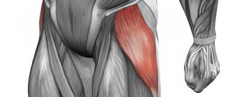
The device of the adductor muscle of the thigh
The adductor muscle of the thigh is a complex muscle group that forms the largest fiber stretching along the inner side of the thigh. It pulls the limb to the middle of the body, thanks to its anatomical structure, the hips are flexed, rotated outward (supination) and the legs are brought to the center.
The adductors include:
- Gracilis muscle: stretches from the pubis to the tibia.
- Long and short (the adductor femoris brevis muscle serves to flex the leg. It starts on the pubic bone and runs to the ischial tuberosity, stretches to the medial lip of the femur, and ends at the linea aspera of the femur).
- Big. Its attachment point begins at the seat. The adductor magnus muscle moves the thigh and extends the pelvis.
- Pectineus muscle: extends from the pelvis.
Each of them participates in the formation of a common tendon in the terminal part of the thigh.
If you have not yet formed an understanding of what was said above, you can simplify the diagram.
If you want to lose weight and are working on your inner or outer thighs in isolation, don’t expect quick results!
The most effective exercises for the thighs and buttocks, both in the gym and at home, are those that dynamically engage the entire lower body muscle group through a full range of motion. Hip abduction or adduction machines are a waste of time!
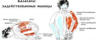
Contraction of the ears reduces the traction of the vertebrae. Therefore, prolonged contraction causes the vertebrae to crush the discs. If we don't resolve this constant tension, it can cause low back pain, sciatica, pinching, dislocated bulges and include hernia.
In addition, if this contraction only shortens, then the torsion bar can cause acquired scoliosis. If the compression is bilateral, it causes lumbar hyperlordosis. For these reasons, it is important to stretch it regularly. Pathology of sedentary and athletes.
One of the reasons behind the reduction of psoas is long hours of work: people who have official jobs or drivers are ideal candidates suffering from this problem. On the other hand, cyclists, medical practitioners and runners are the athletes with the most ballots to be subject to cuts. This is due to the movement performed in these sports: in all of them, the movement of the hip is the main movement.
Don't fall victim to deception and inaccurate information in fitness magazines. If you want your thighs and buttocks to be in the best possible shape, follow a program of full-body training, a balanced diet and a set of cardiovascular exercises.
Bone structure
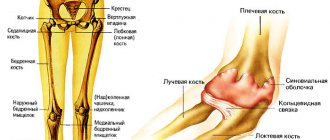
At the heart of this part of the limb is a strong femur surrounded by powerful muscles. This part of the skeleton is equal to a quarter of human height. In structure, it resembles an elongated tube, expanding at both ends, inside which is yellow bone marrow. At the top there is a round head, connected to the body of the bone by the neck. At the junction there are two tubercles - the greater and lesser trochanters, which are necessary for the attachment of muscle fibers.
On the lower edge there are two condyles with epicondyles - lateral and medial. They are necessary for securing the ligamentous fibers.
The bone surface is covered by a connective tissue layer, which is penetrated by nerve endings and vascular networks. It's called the periosteum. Its inner layer contains stem cells. They promote the growth of skeletal tissues and the healing of cracks and fractures.
The body of the bone itself consists of mineral tubular tissue; it is quite rigid and dense. At the ends it transforms into a spongy structure reminiscent of pumice. She knows how to gradually “adapt” to changes when walking, playing sports, or wearing heels. The full structure of the bone can be seen in the photo.
Femur
The femur is the largest tubular bone. Its body is cylindrical and slightly curved anteriorly; a rough line stretches along its posterior surface, which serves for muscle attachment. The body expands downwards. On the proximalproximal [eg. end, phalanx] - (proximalis) - a point on a limb (edge of a bone or muscle) or an entire structure (phalanx, muscle), less distant from the body.
Antonym - distal...click for details..epiphysis-epiphysis (from the Greek epiphysis - growth, lump) - 1) pineal gland, pineal gland, an organ of vertebrates and humans, located between the anterior tubercles of the quadrigeminal cerebrum and connected through a pedicle with the 3rd ventricle 2)…
click for details.. there is the head of the femur, which has an articular surface that serves to articulate with the acetabulum. There is a pit in the middle of the surface of the head. The head is connected to the body of the bone by a well-defined neck, the axis of which in relation to the longitudinal axis of the body of the femur is located at approximately an angle of 130°.
At the point where the neck meets the body, there are two tubercles: the greater trochanter and the lesser trochanter. The first protrudes is the lateral-lateral [edge] - lateralis - the side lying further from the median (central) plane, i.e. outer side. Antonym - medial edge. ...click for details..., easily palpable under the skin;
Read more: Arm training for mass and relief for men: 8 week specialization program for arms in the gym
Distaldistal[eg. end, phalanx] (distalis) - the end of a muscle or bone of a limb or an entire structure (phalanx, muscle) most distant from the body. Antonym - proximal....click for details.. the end of the body of the femur, expanding, without a sharp border, passes into two condyles - medialmedial[edge] - medialis - the side lying closer to the median (central) plane, i.e.
inner side. Antonym - lateral edge. ...click for details.. and lateral, between which there is an intercondylar fossa, clearly visible from behind. The femoral condyles have articular surfaces that serve for articulation with the tibia and the patella. The radius of the surface of the condyles (if you look at them in profile) decreases posteriorly, which gives the contour of the condyles the shape of a segment of a spiral.
Spasm of the hip adductors in infants. Diagnostics
How can you tell if a newborn baby has adductor spasm? Since the adductors are adductor muscles, they will resist when stretched, i.e. To detect tension, the legs need to be spread. This is done with the child lying on his back carefully and slowly so as not to cause pain to the baby. The child’s knees can be bent, which is more comfortable, and tension on the shin flexors is useless, because they are also partially involved in hip adduction. To be sure, you need to do the spreading several times, because children can sometimes simply actively resist and not allow their legs to separate. By the way, the child should be calm at this moment and not cry.
Hip bones
This area contains the large femur. It is presented in the form of a cylinder, there is a head at the upper end, the greater and lesser trochanters are located outside, and muscle fibers are attached to them. Posteriorly there is an intertrochanteric ridge.
The beginning of the bone is connected to the hip joint. The lower (distal) end is expanded, forms a pair of processes - the lateral and medial condyles, the zone of attachment of muscles and ligaments.
The structure of the bone and its massiveness are due to the fact that it bears the main load to support the body.
Fascia, ligaments, joints
The thigh is covered by the fascia lata, which is divided in Scarp's triangle into:
- deep;
- superficial.
The first has a loose structure, lies among the muscle fibers and carries lymphatic and blood vessels and nerves. The second is dense and durable, enveloping the thigh from the outside.
The hip joint is supported by ligaments:
- iliofemoral;
- ischiofemoral;
- pubofemoral.
These elements ensure the stability of the joint and prevent it from bending and injury during movement.
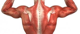
Muscles
The hip can be moved by a group of muscles. They envelop the thigh on all sides and thereby allow it to perform flexion-extension and rotational movements. Anatomy suggests the presence of the following muscles:
- The quadriceps are the largest in the human body. It is called so because it consists of 4 heads, which are attached to the patella and can be easily felt under the skin. The main function is to flex the hip, shin and knee joints.
- The tailor is the longest muscle in the human body. Has the shape of a spiral. Thanks to it, the leg can bend at the knee joint.
- Comb and tender - located on the inside. Needed to bend the lower leg. They also supinate the hip during movement.
- Biceps - this muscle is responsible for flexing the tibia at the knee joint and raising the leg when the knee is extended.
- Semitendinosus - one third consists of tendons and its responsibilities coincide with the biceps femoris muscle.
- Semimembranosus - is used during circular movements of the lower leg.
- Popliteal - pulls back the cartilaginous capsule when bending the knee joint.
The muscles of the lower extremities, due to constantly undergoing high loads, are among the most powerful in the human body.
Skeletal anatomy
With a system as large as the legs, knowing just the muscles is not enough. Learn more about the bones and joints you also need to walk, run and squat!
- Pelvis
The basin has the shape of a bowl. It binds the lower body together and performs two basic movements: anterior pelvic tilt and posterior pelvic tilt.
- Hip joint
The hip joint is where the thigh bone connects to the pelvis. This is a kind of hinge, thanks to which you can bend and extend your legs, adduct and abduct your hip, and rotate.
- Knees
The knee joint connects the femur, tibia and patella. It does more than just help you do flexion, extension and rotation. The knee is critical in every leg exercise you do.
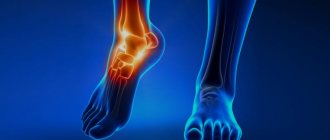
- Ankles (ankles)
The ankles control two basic movements: plantar flexion—curling the toes down toward the sole—and forward dorsiflexion of the foot.
Key Exercises
Today you have read a lot about the structure of your body and muscle functions, all that remains is to understand what to do in the gym. Below are effective exercises that will help you strengthen your leg muscles and achieve a balanced physique.
Exercise 1. Front squats.
- Their advantage is that almost all leg muscles are involved in the work. As you lower, you engage your quadriceps, biceps, and glutes. And when you get up - knee and quadriceps. If you want to choose one exercise to start developing your legs, front squats are great. You will get a good return on your investment. Hold the barbell high on your chest, almost at the bottom of your throat. It's uncomfortable, but it really is the best position. Feet shoulder-width apart, toes slightly outward. Distribute your body weight approximately in the middle of your feet. Sit parallel or lower, and then return to the starting position.
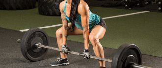
Exercise 2. Romanian deadlift.
- This is a fantastic exercise that isolates your glutes and hamstrings. Focus on pushing your hips back. Keep your knees soft and your spine in a natural curve. As you push your hips back, your glutes and hamstrings stretch. Complete the movement by thrusting your pelvis forward and squeezing your glutes. A full range of motion will give you the challenge you're looking for.
Exercise 3. Lunges.
- Training on one leg puts stress on the muscles and forces the body to stabilize. Keep your torso straight and nice and step forward with one leg. Push off and return to full range of motion. Lunges work every muscle in the leg, including the quads, biceps, glutes, gluteus minimus, and hip adductors.
Exercise 4. Standing calf raises.
- You can use your own weight, dumbbells or barbell. The great thing about this exercise is that it is difficult to do incorrectly. Keep your knees nice and straight to stretch your Achilles tendon and calf muscles. At the bottom of the movement, pause for a second or two.
Major diseases
In addition to injuries to soft tissues and bones, pain is often caused by various processes in the bones. Sometimes the pain radiates to the thigh due to pathologies of the spine (osteochondrosis, spondylosis). To find out the cause of the pain, it is necessary to observe the nature of the painful sensations, their intensity, as well as the reaction to the load on the hip and changes in the position of the limb. Pain in the hip can be sharp, dull, aching, cutting - depending on the situation.
Soft tissue injuries
Mechanical damage is the most common cause of pain in the hips . Impacts and mechanical injuries refer to damage to the soft tissues of the thigh, accompanied by ruptures of blood vessels and nerve fibers. In this case, the skin can remain intact, while an area of hemorrhage forms under it.
Soft tissue bruise of the thigh
The injury occurs as a result of falls or blows. This diagnosis is characterized by the following features:
- type of pain - dull, aching, aggravated by pressing on the damaged surface, the motor ability of the limb is preserved;
- localization of pain - one-sided, at the site of injury;
- additional symptoms include the formation of a hematoma (a blue-violet, irregularly shaped area that appears as a result of the rupture of small blood vessels under the skin).
A bruise is diagnosed during an examination, and sometimes an x-ray is taken to rule out a fracture. If the bone is intact and there is a hematoma, the doctor diagnoses “bruise of the soft tissues of the thigh.” In most cases treatment of a bruise is not required, since healing of damaged tissue occurs on its own without the need for outside help. But in some cases, the help of a surgeon or traumatologist is required if the injury is severe and an extensive hematoma has formed in its place. In this case, a large volume of blood in the subcutaneous and intermuscular space can compress neighboring nerves, causing pain. The doctor opens the hematoma with a medical instrument and removes the blood.
Ask your question to a neurologist for free Irina Martynova. Graduated from Voronezh State Medical University named after. N.N. Burdenko. Clinical resident and neurologist BUZ VO \"Moscow Polyclinic\".Ask a question>>
Hip sprain
Sprained hip ligaments is a complete or partial rupture of small fibers of ligamentous tissue , which occurs as a result of disproportionate physical activity (during sports, lifting weights), falls, slips, sudden changes in body position or heavy load without prior preparation (warm-up). Children and adolescents with underdeveloped muscle structures, as well as older people with osteoporosis, are most often susceptible to such injuries.
Main signs of sprain:
- type of pain - acute, intensifying when trying to move the leg;
- localization of pain - in the hip joint, one-sided, over time “spreads” along the thigh towards the lower leg, less often radiates to the lower back;
- additional symptoms are swelling at the site of injury, hyperemia of the skin over the injured area.
Sprain of the hip ligaments is diagnosed during examination and palpation. An orthopedic doctor or traumatologist moves the patient’s limb in different directions and asks the patient to perform simple exercises and, depending on the success of their implementation, makes a preliminary diagnosis. The final diagnosis is made using an x-ray, which usually shows joint deformity.
Treatment of injury involves applying a fixing bandage that limits the mobility of the limb. Further therapy depends on the degree of ligament damage. With the relative preservation of the integrity of the ligamentous tissues, conservative treatment is carried out (taking anti-inflammatory and analgesic medications, ensuring rest). As the ligaments are restored, exercise therapy is prescribed, aimed at restoring functionality to the joint. For complete ligament rupture and/or avulsion fracture, surgery is performed.
Bone injuries
Fractures are another cause of hip pain . They also occur as a result of rough mechanical impact - shocks, falls, sudden compression, improper load distribution and other factors.
Femoral neck fracture
Often pain occurs due to a hip fracture, especially in people over 65 years of age .
Aging is usually accompanied by osteoporosis - increased fragility of bones, and even with light loads the integrity of the bone can be compromised. Typically a fracture occurs as a result of a fall. The symptoms of a fracture are as follows:
- the nature of the pain is acute;
- localization of pain - in the upper thigh with irradiation to the groin;
- additional symptoms are rotation of the foot outward relative to the knee, limited mobility of the leg, inability to walk and stand.
is diagnosed using radiography and MRI of the joint. You can also determine a femoral neck fracture by tapping or pressing on the heel: the patient will experience unpleasant and even painful sensations.
Treatment of a hip fracture can be quite difficult, especially in old age. Plaster application does not have an effect, so the victim is prescribed surgical intervention - osteosynthesis (fixation of joint fragments with metal screws), as well as endoprosthetics (full or partial joint replacement).
Pertrochanteric femoral fracture
This type of fracture is also most common in women over 65 years of age and occurs as a result of falling on the side (while walking on a slippery surface in winter, with sudden movements).
This diagnosis has the following symptoms:
- the nature of the pain is strong, very sharp;
- localization - in the area of injury in the upper thigh;
- additional symptoms are “stuck heel syndrome,” in which the patient cannot lift an outstretched leg while lying on his back.
Accurate diagnosis is only possible based on radiography. Treatment of a pertrochanteric fracture today is practiced in the form of surgery, in which the bone is pinned and fixed in the correct position. The operation allows you to quickly recover from injury, and the procedure itself is minimally invasive (a small incision is made) and lasts about 20 minutes.
Soft tissue inflammation
Often the thighs on the outside hurt not because of mechanical damage, but because of the inflammatory process occurring in the soft tissues.
Myositis
One of the causes of pain in the soft tissues of the thigh is myositis , which occurs due to hypothermia, injury, infectious or autoimmune processes, when the body begins to perceive tissue cells as foreign and attack them. The patient feels pain of moderate intensity due to weakening of the thigh muscles .
is diagnosed based on a survey, examination, and a blood test that detects eosinophilic leukocytosis. A soft tissue biopsy is also performed.
Treatment of myositis is complex:
- ensuring rest (bed rest);
- diet correction (strengthening the diet with vitamins and mineral complexes).
Depending on the cause of the disease, treatment is carried out with antibiotics (for infection), immunosuppressants and glucocorticosteroids (for an autoimmune cause), non-steroidal anti-inflammatory drugs, physiotherapy and massage (if the doctor allows).
Trochanteritis
Trochanteritis is an inflammation of the tendons that connect the lesser and greater trochanters to the femur . Most often, the pathological process occurs due to injuries, due to hypothermia or overload. The pain is aching, pressing, aggravated by exertion (walking, climbing stairs), hypothermia. Localization of unpleasant sensations is in the outer side part (“breeches”).
diagnosed through examination and questioning, blood tests, X-rays or MRIs of the hip.
Treatment is conservative and involves the use of non-steroidal drugs. In more complex cases, injections of glucocorticosteroids into the tendon area are prescribed, which are done once every 2 weeks. Physical therapy is also prescribed, less often laser therapy, massage with rubbing of anti-inflammatory ointments.
Inflammatory bone lesion
The bones and joints of the hip are also susceptible to negative factors leading to pathological processes that cause pain.
Coxarthrosis
The main symptom of coxarthrosis is pain in the groin, radiating to the outer front and side of the thigh, and less often to the buttock and knee. Both joints or just one can hurt. It becomes difficult for the patient to move the limb, especially to move it to the side. A crunching sound is heard in the joint, and the leg may look slightly shorter than the other.
Coxarthrosis is diagnosed using radiography - the image shows an increase in the neck-diaphyseal angle, dysplasia or changes in the proximal part of the femur.
Treatment of the disease:
- conservative, at an early stage - with the help of anti-inflammatory drugs, chondroprotectors, intra-articular steroid injections, warming ointments;
- surgical - if the hip joint is severely damaged, endoprosthetics (joint replacement) is performed.
Aseptic necrosis
Aseptic necrosis is very similar in symptoms to coxarthrosis, but is characterized by high intensity pain , which becomes unbearable as the pathological process develops. The disease begins due to the cessation of blood supply to this part of the joint; the process itself proceeds quickly and is accompanied by severe night pain. Characteristic for this disease is the age of the patients: most often it affects men from 20 to 45 years old, while women are 5-6 times less likely to suffer from it.
Diagnosis of hip joint disease is carried out using modern research methods - X-rays and MRI. An experienced doctor can make a diagnosis based on symptoms and examination of the limb, but ultimately everything is decided by an X-ray examination of the joint and bone.
Therapy consists of restoring nutrition to the femoral head. Non-steroidal and steroidal agents, chondroprotectors and calcium preparations are also used to accelerate the restoration of damaged bone tissue.
Expert opinion
Prikhodko Arkady Arkadievich
Rheumatologist - city clinic, Moscow. Education: FGBNU NIIR named after V.A. Nasonova, Astrakhan State Medical Academy.
Sometimes pain on the outside of the thigh can be caused by cancer, pathologies of the arteries and veins. In case of diseases of the spine, unpleasant sensations on the outside of the thighs may be reflected, but we will not dwell on these reasons in detail, since we have already discussed them in the article “Pain on the inside of the thigh.”
Nervous structure
The nerve endings of the lower extremities descend from the lumbosacral plexus.
Their function is to transmit signals from the central nervous system and back to enable the muscles to move the limb correctly. They also allow the skin to sense touch and temperature changes. If there are disturbances in this area, a person begins to have problems with the muscles of the thigh, flexion and extension of the knees. The main nerve passing through the pelvis along the posterior and outer regions of the femur has a similar name. Its branches provide communication with the central nervous system of almost all organs and tissues of the upper leg. Peripheral nerves branch from the main trunk:
- subcutaneous;
- internal musculocutaneous;
- lateral and anterior cutaneous;
- median muscular.
The obturator nerve, which runs from the lumbar plexus along the lateral wall of the pelvis, also plays an important role. It diverges into two branches - articular and muscular, which connect the corresponding structures near the obturator canal with the central nervous system.
The corresponding part of the genital femoral nerve innervates the oblique and transverse muscles in the inner thigh and the skin near Scarpa's triangle.
The sciatic and posterior cutaneous nerves depart from the sacral plexus.
The first of them, with the help of lateral branches, innervates the muscular tissues of the dorsal surface of the thigh, participating in flexion of the knee joint. Additionally, it transmits signals to the fibers of the midfemoral region, assisting its adductor actions. The sciatic nerve ends with two large branches - the common peroneal and tibial.
The second, with the help of auxiliary branches, creates the conditions for motor innervation of muscle tissue behind the lower leg. Through its actions, it promotes extension of the ankle joint and flexion of the toes. Responsible for their motor function are two nerve endings located in the sole of the foot.
The common peroneal branch innervates the corresponding muscles, as well as the ventral tissues of the leg, which allows free flexion and lateral movement of the ankle. The influence of this branch is also responsible for the extension of the fingers.
The posterior cutaneous branch participates in the motor innervation of the pelvis, creating conditions for the work of the gluteus maximus muscle. In addition, its activity helps abduct the femoral joint and provides sensitivity to the dorsal femoral surface and the top of the ankle joint.
Diseases of muscle tissue, blood vessels, bones, and nerves of the thigh are not uncommon. Knowledge of the anatomical structure and the use of modern hardware diagnostic techniques allows us to identify them at an early stage, avoiding complications and disability.
Source: NogoStop.ru
Blood and lymphatic vessels
Many vessels pass through the femoral part, each feeding specific organs and structures. The most important is the femoral artery (in Latin - a. femoralis). It continues the iliac vessel, descends along the anterior outer part of the thigh through the vascular lacuna into the popliteal cavity, where it transforms into the artery of the same name. In Scarp's triangle, the main vessel of the femur is covered only by connective tissue and skin. Other femoral arteries depart from it:
- superficial;
- deep;
- superficial epigastric;
- medial;
- lateral;
- perforating;
- external genitalia;
- descending knee
The femoral vein arises from the azygos popliteal vein and has about eight peripheral branches. One of them is the deep vein, which “works” on the back of the thigh. Also, large venous vessels pass medially and laterally and serve the corresponding parts of the upper limb. The superficial circulatory network is located directly under the skin.
In the femoral region there are large lymph nodes - superficial and deep inguinal. The first are located under the skin on a wide connective tissue element along the inguinal fold and on its anterolateral surface. You can really feel them with your fingers. The second ones are located deep in the thigh near the vein. The largest is located directly at the vascular lacuna.
Additional small lymph nodes are located singly and in groups in different femoral sections along the lymphatic vessels.
The latter also vary in depth. Superficial vessels go from the wall of the peritoneum and genital organs to the lymph nodes, and deep ones - from the lymphocapillaries of the muscles, joints, and bone structures. The lymph nodes of the femoral part connected by a vascular network form the inguinal lymphatic plexus. A complete diagram of the vessels can be seen in the photo.
Fascia and ligaments
Fascia is a sheath of connective tissue that covers organs, blood vessels, nerves and forms sheaths for muscles. In the hip area, you can distinguish the fascia lata, which is the thickest in the human body. In terms of strength, it is not inferior to the tendon bundle, especially in the area of the middle part of the thigh. In the area of Scarp's triangle, it is divided into two plates: superficial (subcutaneous) and deep. The subcutaneous tissue loses density and becomes loose, as saphenous veins, lymphatic vessels, nerves, and adipose tissue pass through it.
The hip joint capsule is strengthened by a powerful ligamentous system. In front it is the iliofemoral and pubofemoral ligament, in the back it is the ischiofemoral ligament.
Meet the femoral region
The human thigh, or femoral region, is the part of the leg from the oblique fold of skin in the groin area to the knee joint. It is worth recalling this because in fiction, for some reason, “hips” are sometimes called buttocks. The hip performs an important supporting function. It allows us to stay upright. And also walk, run, jump, crawl...
The hip frame is represented by a strong femur. It is surrounded by powerful muscles. The muscles give the thigh volume, elasticity and allow us to move our legs. The outside of the thigh is enveloped in subcutaneous fat and skin.
All layers of the femoral region are penetrated by blood and lymphatic vessels and nerve endings. Without these “communications” not a single organ of the human body can function. Large arteries, veins and nerve trunks are located in the depths, near the femur. They are protected from damage by muscles and subcutaneous tissue. Small branches of blood vessels and nerves penetrate all the tissues of the femoral region.
The proximal part of the femur (closer to the head, upper) forms a movable connection with the pelvic bone, and its lower part connects to the bones of the lower leg in a complex knee joint.
What is the hip and what functions does it perform?
The thigh is the upper part of the lower limb, the area between the pelvis and the knee. It is a kind of connecting link between the lower limbs and the torso.
Reference . “Femur” is the name of the thigh in Latin.
The large femoral region of the leg is responsible for the following functions :
- flexion-extension of the leg;
- rotation of the limb;
- abduction-adduction;
- moving, turning, braking;
- participation in the process of hematopoiesis (blood cells form, develop and mature in the bone marrow: leukocytes, erythrocytes, platelets).
This part of the leg serves as a support for the body , providing stability to the human body.