Basic anatomy of the hip joint: bones that form the joint
The human hip joint is formed by two bones, the surfaces of which ideally coincide, like pieces of a puzzle. The acetabulum on the surface of the ilium plays the role of a kind of pocket into which the spherical process of the femur is immersed - the head, completely covered with durable and elastic cartilage.
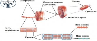
Soft and painless sliding between two fairly tightly adjacent bones is achieved due to the special structure of cartilaginous tissue. The combination of collagen and elastin fibers allows you to maintain a rigid and at the same time elastic structure of cartilage, and proteoglycan molecules and the water included in the composition guarantee the necessary pliability and elasticity.
https://www.youtube.com/watch?v=rnu3FyA3R3o
The joint cavity is limited by a special capsule, the basis of which is fibrous fibers. These molecules are characterized by increased strength, so that even under great pressure the joint retains its integrity and original shape. However, this reserve is not unlimited, and, unfortunately, it is impossible to guarantee 100% the impossibility of dislocation: with inadequate loads, strong external pressure or a sharp displacement in space, such an atypical injury is quite real.
Musculoskeletal system of the foot
Ligaments play a very important role in the functionality of the hip joint. It is these super-strong fibers that maintain the optimal shape of the joint, ensure adequate mobility and activity of the joint, and protect against injury and deformation. The ligamentous apparatus of the hip joint is represented by the most powerful fibers:
- The iliofemoral ligament is the most powerful and durable ligament in the human body, capable of withstanding incredible loads without tears or sprains. Experimental experiments have shown that its fibers are capable of withstanding a load comparable to the weight of 3 centners. It is thanks to this that the joint remains protected during intense training, unsuccessful movements and other unpleasant surprises that affect the mobility of the femoral joint.
- The ischiofemoral ligament is a much thinner and softer ligament that controls the degree of pronation of the femur. It is, as it were, woven into the articular capsule, located from the ischium all the way to the trochanteric fossa.
- The pubofemoral ligament is responsible for the abduction angle of the free femur of the lower limb. Its fibers, like the ischiofemoral ligament, penetrate into the articular capsule, however, they originate not at the ischium, but at the pubic joint.
- The circular ligament does not leave the joint capsule. As the name suggests, it is located in a circle, encircling the head and neck of the femur in a tight loop and secured to the anterior surface of the lower bone.
- The ligament of the femoral head is the most original in the anatomy of the hip joint. Unlike its “colleagues”, it does not directly protect the joint and does not control its mobility; the functions of this ligament are to preserve the blood vessels with which it is penetrated. This feature is explained by its location, which coincides with the trajectory of the vessels: the ligament begins at the acetabulum and ends at the head of the femur.
Detailed structure
The main structural units of the limbs are joints, bones, muscles, blood vessels, nerve fibers, and ligaments. Separately, it is worth highlighting the hands and feet as important and structurally complex elements of the limbs. Next, we'll take a closer look at each unit of leg and arm anatomy.
Bones
The arm is attached by the collarbone and scapula. The collarbone is located at the top of the sternum, and the shoulder blade is located above the upper ribs on the back side. It then articulates with the humerus bone. The latter holds the remaining bones of the arm. The next part is called the forearm. Consists of the ulna and radius bones, which are connected to form a joint.
The next section is the most complex and multifunctional - the brush. Which in turn is divided into departments:
- wrist - eight bones arranged in two rows. They are part of the wrist joint;
- metacarpal bones - five bones located from the wrist to the phalanges. They hardly move, as they support the fingers;
- phalanges of the fingers - four fingers in their structure have proximal, middle and distal bones. And the thumb consists of two - the main and nail bones.
The leg on top consists of the ischium, ilium and pubic bones of the pelvis. They provide support for the body. At the age of eighteen, these bones fuse and form the acetabulum. The femur bone fits into it, forming the joint. Thanks to it, the leg rotates and supports the weight of a person.
The femur ends at the patella, which fits into the kneecap. Next is the tibia and the tibia. And actually the last element is the foot. There are twenty-six bones in its structure.
Leg bones
The foot is divided into three parts, just like the hand:
- tarsus - talus (connected to the tibia), calcaneus, navicular, lateral, intermediate, medial and cuboid;
- metatarsals are five bones that connect the previous part of the foot with the phalanges of the fingers;
- toes - four toes consist of three phalanges (proximal, middle, distal). The thumb has two bones, the middle one is missing.
Joints
The hand consists of three large joints (shoulder, elbow and wrist), as well as a huge number of small ones. Large joints of the upper limbs:
- The shoulder joint is a ball-shaped connection between the shoulder bone and the shoulder blade. Due to the large articular surface of the scapula, this joint has a large range of motion.
- The elbow joint is formed by three bones (humerus, ulna and radius). It has a block fastening system, due to which it can work on flexion, extension, as well as adduction and abduction.
- The wrist joint is formed by the radius and a number of carpal bones. Movements are possible in three planes.
Shoulder structure
The hand consists of four types of joints:
- midcarpal - formed between two rows of carpal bones;
- carpometacarpal joints;
- metacarpophalangeal - perform the function of holding the phalanges on the stationary area of the hand;
- interphalangeal - four fingers are formed by two joints, and the thumb has one.
The longest range of movements is provided by the last two groups of joints. The remaining joints perform an additional function without particularly participating in movements.
The following is a table of the joints of the human lower extremities.
| Joint name | Short description |
| Hip | The ball and socket joint is the most powerful in the human body and performs a wide range of movements. The joint cannot be felt with your hands, as it is located deep under the muscle tissue. It is formed by the head of the femur and the acetabulum. |
| Knee | The condylar type is formed by three bones - the femur, tibia and patella. Inside the articular cavity there are two semilunar septa made of cartilage tissue. They are called menisci. The articulation contains several bags with synovium, not connected to the main cavity of the joint. The knee can bend, straighten and rotate within a small range. The ligaments are placed crosswise. |
| Ankle | Block joint of the leg and foot. It is formed from two tibia bones (tibia and fibula), which are arranged in the form of a fork and capture the talus bone. The articulation bursa resembles a cuff, which has strong walls on the sides, and the front and back are represented by folds of a loose nature. Functional tasks - flexion and extension. |
| Finger joints | The phalanges, like the hands, have two joints on four fingers and one on the thumb. They are characterized by less mobility than on the hand, since there is no functional need for this. |
Ligaments
Ligaments connect bones to each other, fixing them in a certain position, and limit the range of their movements. Ligaments are located in and around joints, and are named identically to the bone joints to which they are attached.
The following bundles are in your hands:
- three ligaments of the clavicle;
- three articular-brachial (upper, lower and middle);
- four ligaments of the elbow (ulnar, anterior, radial and posterior);
- ligaments in the wrist joint (lateral, radial, ulnar, dorsal, palmar and intercarpal);
- ligament that closes the flexor retinaculum - a special ligament with a special purpose, which is located in the carpal tunnel, prevents pinching of blood vessels and nerves;
- ligaments of the hand - there are many of them, connecting the small bones of the hand.
The lower limbs are represented by the following ligaments:
- hip joint - three longitudinal and one circular;
- knee – tibia and fibula, internal cruciate, popliteal and patellar ligament;
- ankle - external lateral (anterior, posterior and calcaneal fibula), four internal lateral, interosseous (passes through the entire lower leg). There is also a posterior inferior (does not allow the bones to rotate inward), anterior fibular and transverse.
Muscles
The leg muscles are very strong, since their main function is to ensure mobility of bones and joints. On the thigh is the quadriceps, which allows you to bend the lower leg. The sartorius muscle also provides flexion of the tibia and femur. It also allows you to rotate your hip. Two more groups of leg muscles are adductors and medials. Thanks to them, the femur rotates inward, raising and lowering the thigh towards the body.
Leg muscles
The calf muscles allow you to raise and lower your foot. The external muscles of the lower leg lower the foot and flex the sole, and the rear muscles raise the heel and allow you to stand on your toes. There are eleven muscles of different sizes and shapes in the foot. They are responsible for the movements of the fingers and lifting the foot off the surface (in total, thirty-eight different muscles of the body are used to perform this function).
The muscle structure of the arms is very numerous. It is very difficult to list all the names, since the forearm alone has about twenty bundles. It is worth highlighting that there are two types of muscle fibers - extensors at the back of the bones, flexors at the front. They envelop the entire structure of the upper limb and allow it to fulfill its functional purposes. The muscles of the hand are divided into thenars, gopotenars and middle ones.
Blood circulation and nerve fibers
This part of the structure provides the limbs with blood supply (oxygen and micronutrients) and innervation (impulses to the spinal cord and brain and back). The upper limbs are supplied by the subclavian artery, which then passes into the axillary and brachial arteries. And then the branches connect and flow into the ulnar artery, from which there is a branch further along the arm and hand.
The venous network has a similar structure, and there are also subcutaneous vessels on both parts of the arm. The nervous system begins in the shoulder area. And then there is a branching through the nerve columns - axillary, musculocutaneous, radial, median fibers and ulnar.
Blood circulation in the legs also passes in three directions - arteries, veins and capillaries. The latter are small vessels located close to the surface of the skin. They receive blood from the eight large veins of the leg, which in turn connect to form the femoral vein. Arteries pass through the entire leg - femoral, tibial (anterior and posterior), under the knee, dorsal for blood supply to the foot, lateral, medial (the last two on the sole).
The nerve canals in the leg begin from the lumbar region and move to the nerve of the thigh. The innervation of the lower leg is carried out by the perineal, tibial and subcutaneous fibers. Three nerves are responsible for the foot - medial, lateral and gastrocnemius.
All nerve channels are interconnected and impulses pass from one to another. This system provokes referred pain. When the injury is localized in one place, and the pain syndrome spreads to several parts of the limb. Or there is a problem in the spine, but the leg hurts.
Vascular and nervous systems of the foot
Like any joint of the human body, the hip joint is not distinguished by a high organization of the nervous system: the endings localized in this area mainly innervate the muscle fibers, regulating the degree of sensitivity and coordinated work of each muscle group in response to external influences. Conventionally, all nerve fibers of the hip region can be divided into 3 groups:
- anterior external, which includes branches of the femoral nerve;
- anterior internal - branches of the obturator nerve;
- posterior - branches of the sciatic nerve.
Each group is localized in a specific area of the thigh, for which it is responsible in the complex structure of the nervous system of the body in general and the lower extremities in particular.
To allow blood to flow to the feet, there are tibial arteries in front and behind. They stretch along the very foot on the sole. Small connections and circles extend from these large arteries.
The veins on the back side are responsible for the outflow. They appear intertwined and supply blood to the greater and lesser saphenous veins in the lower leg.
They form an integral part of the normal functioning of the human foot. They are responsible for sensations:
- - pain;
- — vibrations;
- - touch;
- - cold or heat.
Nerve signals leaving the central nervous system along the sural, peroneal, superficial and tibial nerves reach the spinal cord and are processed there.
Nerves transmit signals to muscles, being essentially reflexes - voluntary or involuntary (independent of human will). Involuntary ones include the work of the glands (sebaceous and sweat), vascular tone.
As for the skin, there are several zones on the foot that differ in density, structure, and elasticity. For example, the sole leather is high density and the heel leather is thick. Initially, the skin of the palms and feet is the same, but over time and with increasing loads, additional layers appear. The dorsum of the foot is smooth and elastic, with nerve endings.
Drawing a conclusion, we can say that nature has done everything to ensure that the foot can withstand enormous pressure.
Functional purpose
The main functions of the foot are:
- Spring . This is done due to the presence of arches and their ability to act as a shock absorber. Thanks to the spring function, the feet absorb up to 80% of the energy when touching the support. This ensures that you can run, walk, and jump without permanent damage.
- Reflexogenic . The activity of the nervous system, which continuously monitors the location of the body. A small area of the foot contains many functional points through which the foot is connected to the brain and organs.
- Balancing . The joints are responsible for it. They guarantee the ability to move while maintaining position.
- Jogging . With the help of the pushing function, a person moves forward: the foot receives kinetic energy in the instant of contact with the support, restrains it during the roll from heel to toe, returns it back to the body, lifting off for a new swing.
The primary specialization is maintaining the weight of the body and ensuring its movement in space. The human foot has 3 points of bone support: 2 are located in the frontal region, 1 in the posterior. When walking on your toes, stability depends on the length of your toes.
The front part of the foot, especially in the toes, is mobile and compressible. Based on the position of the anterior section relative to the posterior one, the feet can be divided into straight, adducted and abducted. Also, the foot has the ability to twist along the longitudinal axis, the outer and inner edges to rise.
Anatomical structure of the human leg
Functions
The leg has many functions:
- walking;
- run;
- jumping;
- crawl;
- swimming;
- support, etc.
If you remember the anatomy, the leg has three parts - thigh, lower leg, foot.
The musculature of the hip joint is represented by fibers of various types and functionality. This is primarily due to the varied trajectory of movement that the hip can perform. So, if we classify muscle fibers into groups according to function, in the anatomy of the hip joint we should highlight:
- The transverse, or frontal, muscle group, which is responsible for flexion and extension of the lower limb in the pelvic area. Among them are the flexor muscles (sartorius, iliopsoas, pectineus, rectus, tensor fascia lata) and hip extensor muscles (gluteus maximus, adductor magnus, semitendinosus, semimembranosus and biceps). Thanks to their coordinated work, a person can sit down and stand up, squat down and take a vertical position, pull his legs to his chest and straighten up.
- The anteroposterior, or sagittal, muscles regulate the adduction and abduction of the leg. This group includes adductors (major, short and long adductors, gracilis and pectineus) and abductors (obturator internus, tensor fascia lata, twin, piriformis, gluteus medius and minimus) muscle fibers.
- The longitudinal muscle group coordinates the rotation of the hip. Here the supinator muscles are distinguished (gemini, piriformis, iliopsoas, quadratus, sartorius, obturator, gluteus maximus and posterior groups of the middle and small gluteal fibers) and pronators (tensor fascia lata, semitendinosus, semimembranosus, anterior group of the middle and small gluteal fibers) .
Each of the muscles represented in the anatomy of the hip joint performs not only a motor function: powerful fibers take on part of the load during movements. And the more trained they are, the better they cope with pressure, thereby unloading the joint and performing a shock-absorbing function. Thanks to this, the likelihood of injury due to unsuccessful movements is also reduced, since the muscles are more mobile and extensible than the tissues of the joint.
Proper leg training
Rule No. 1 – priority of basic exercises; Rule No. 2 – progression of loads; Rule No. 3 – alternating training for fast and slow muscle fibers; Rule No. 4 – we take out our legs on a separate training day; Rule No. 5 – the load always accumulates in the target muscle group, so we be sure to develop muscle sense ; Rule No. 6 – keep a training diary and experiment with the effectiveness of different training schemes; Rule No. 7 – always control the weight, that is, the negative phase in any exercise lasts at least three seconds; Rule No. 8 – we always train the lagging muscle group first .
The training scheme for slow muscle fibers assumes a large CP, that is, such training should be volumetric and high-intensity. It may be useful to use a 3 to 20 scheme, when 3 sets of 20 repetitions are performed, and for the triceps muscle the number of repetitions can reach 35-40.
The training scheme for fast muscle fibers is a variant of more strength training, when the athlete performs sets of 3-5 repetitions and rests for a long time between them. During such training, attention should be paid only to large muscle groups, and they should be trained exclusively with basic exercises.
Diagnosis of diseases
The foot is regularly exposed to loads, either static or shock. Injuries are a common occurrence for her. They are almost always accompanied by pain, enlargement of some epiphyses, swelling, and curvature. Pathology can be detected by x-ray.
Arthrosis
This is a disease during which cartilage loses its elasticity. This often disrupts metabolic processes. Pain, crunching, swelling appears.
Causes of arthrosis:
- - infectious diseases;
- - allergies;
- - systemic diseases - lupus erythematosus, scleroderma;
- - tuberculosis;
- - syphilis;
- - dislocation or bruise.
The disease develops in 3 stages:
- Pain occurs at first, but goes away after rest. Sometimes deviation of the thumb becomes noticeable. There is a crunching sound when moving.
- Painkillers and anti-inflammatory drugs are taken to dull the pain. The toe becomes severely bent and it becomes impossible to pick up shoes.
- The pain does not go away even when taking analgesics. The deformity spreads to the foot, causing problems with walking.
Arthrosis also greatly affects the ankle, deforming the joint and affecting the cartilage.
This disease can be treated conservatively only at an early stage. Then you will need surgical intervention - endoprosthetics, resection, arthroplasty.
Flat feet
There are congenital or acquired flat feet. Reasons for appearance:
- - excess weight;
- — heavy loads;
- - diseases of nerve endings;
- - injuries;
- - wrong shoes;
- - history of rickets or osteoporosis.
Flat feet exist in two types:
- Transverse - with a decrease in the height of the arch, when the heads of the metatarsal bones contact the ground.
- Longitudinal - that is, the entire foot has contact with the ground. Increased leg fatigue and pain.
Arthritis
A joint disease that affects the entire human body. There are primary and secondary arthritis. The reasons for its appearance are the same as for arthrosis. Symptoms include:
- - pain;
- - leg deformity;
- - swelling, redness;
- - fever, rash, fatigue.
Treatment methods depend on the root cause of the disease and can be physiotherapeutic, medicinal, manual, etc.
Clubfoot
As a rule, it occurs from birth. The cause is a subluxation of the ankle joint. Acquired clubfoot becomes a consequence of trauma to the lower extremities, paralysis, and paresis.
The ankle joint may be damaged or defective. To identify the problem, a diagnostic procedure is prescribed. It may consist of:
- Ultrasound. This diagnostic method is rarely used due to the small size of the ankle joint. But, it allows you to detect a foreign body, swelling due to the accumulation of blood in the joint capsule and view the ligaments.
- Arthroscopy. A minimally invasive method that performs diagnostics by introducing a video camera into the capsule.
- X-ray. The most economical way. It is allowed to take pictures in different projections. Able to detect tumor, fracture, dislocation and other processes.
- MRI. The best type of diagnosis for the condition of the Achilles tendon, ligaments, cartilage. Expensive, but very effective.
- CT scan. Helps assess the condition of the joint. It is considered the most reliable test for arthrosis, neoplasms and fractures.
Examination of bones and joints of the lower extremities
When the first symptoms of lower extremity injuries appear, immediate diagnosis should be carried out to identify the problem at an early stage.
The first symptoms may be:
- the appearance of nagging pain in the calf muscles;
- general weakness of the legs;
- nervous spasms;
- constant hardening of various muscles.
Moreover, if there is even slight pain on a constant basis, this also indicates possible damage or illness.
General inspection
The doctor checks the lower extremities for visual abnormalities (enlarged kneecap, tumors, bruises, blood clots, etc.). The specialist asks the patient to perform some exercises and tell him if pain is felt. In this way, the area where the disease may occur is identified.
Goniometry
Goniometry is an additional examination of the lower extremities using modern technologies. This method allows you to identify deviations in the amplitude of vibration of the joints and patella. That is, if there is any difference from the norm, there is reason to think about it and begin to conduct further research.
Radiation diagnostics of the lower extremities
There are several types of radiation diagnostics:
- X-ray. An image is taken to help replace skeletal damage. However, you should not think that X-rays reveal only cracks and fractures; in some cases, you can notice cavities, a problem associated with a lack of calcium in the body.
- Artography is similar to the previous method, however, images are taken pointwise in the area of the knee joint to check the integrity of the menisci.
- Computed tomography is a modern and expensive method, but extremely effective, because the measurement accuracy error is only a millimeter.
- Radionuclide methods . They help the specialist identify pathologies in the lower extremities and joints.
There are additional research methods prescribed privately:
- ultrasound examination (ultrasound);
- magnetic resonance imaging (MRI).
However, despite the effectiveness of some methods, the most reliable solution is to combine several in order to minimize the possibility of not noticing an illness or injury.
Physiological capabilities of the hip joint
The hip joint can perform movements in three planes at once - frontal, sagittal and vertical. Thanks to the structure of the joint, thought out by nature, a person can easily bend and straighten the hip, move it to the side and bring it to its original position, rotate it in all directions, and at a fairly noticeable angle, the magnitude of which can vary depending on the anatomical features and training of the ligamentous apparatus.
But that’s not all: the hip joint is one of the few joints that can move from the frontal to the sagittal axis, providing the free limb with full circular motion. It is on this ability that a person’s mobility, his physical characteristics and abilities for certain sports (for example, gymnastics, athletics, aerobics, etc.) primarily depend.
The other side of the coin is the rapid wear and tear of the cartilaginous surfaces of the hip joint. The pelvic and femur bones bear the maximum load during walking, running and other types of physical activity, respectively, this pressure is transferred to the joints. The situation can be aggravated by excessively high weight, too intense physical activity, or, conversely, a passive lifestyle in which the muscular system practically does not protect the joint from deformation.
As a result, the cartilage surfaces begin to wear out, become inflamed and become thinner, pain appears, and the trajectory of movement is significantly limited. Even the slightest deviation in the condition of the muscles, ligaments or bones of the hip joint can lead to serious pathology, which subsequently requires long-term and intensive treatment.
However, restoration of the full function of the joint is not always possible: in some cases, surgical intervention is required, in which the affected tissue is replaced with a prosthesis. To prevent this from happening, it is worth monitoring the condition of the musculoskeletal system from a young age, strengthening joints, training the muscle frame wisely and moderately, and taking care of proper and nutritious nutrition of the body. Only in this way can you protect your joints from destruction, and yourself from pain, stiffness of movement and tedious treatment!
Muscle groups
The study of the anatomy and muscular structure of the human body shows that the leg muscles account for up to 40-45% of the mass of all muscle groups. One of the main ones is the gluteal. It includes 3 muscles:
- gluteus minimus, which provides abduction of the lower limb to the side;
- the gluteal medius, which allows you to flex and extend your leg. In addition, it allows a person to stand while bending their body forward;
- gluteus maximus - straightens the leg and ensures a stable position of the torso.