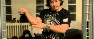
Charles Atlas did not set fantastic records and did not win prestigious titles, which, however, did not prevent him from becoming a cult person in world bodybuilding. His main legacy is a set of isotonic exercises that has withstood a barrage of criticism, but has proven its validity and effectiveness. Our story about the life path and exercise system of Charles Atlas.
ZygoteBody
One of the best services, according to user reviews, is ZygoteBody. This is a resource that allows you to study the structure of the human body using interactive technologies. The previous name of this project was Google Body, which indicates the name of one of the developers. Later the resource was renamed, but the URL and functionality were retained. The interface is designed in English, which is not a problem, since auto-translation can be activated in the browser functions. So, the site loads, and a 3D figure of a woman appears in front of us.
Home page ZygoteBody
Using the mouse, arrows and pointers in the upper left corner of the site, the figure can be enlarged or reduced, as well as rotated in different directions. There is also a high column on the left, moving the slider along which we gradually see all the life support systems of the human body.
Resource functionality
Let's enlarge the picture and click on any organ. A small menu will appear. After clicking on the inverted triangle, information about the selected organ provided by Wikipedia appears. In this menu we will also be able to perform other actions with the image (delete selected organs, move the image to the center of the screen, etc.).
Description of the ventricles of the heart
The table shows the capabilities of ZygoteBody.
| Program versions | Functional |
| Free version | Only viewing of life support systems and their descriptions from Wikipedia is available. |
| Premium | Subscription costs $4 per month. This level of information delivery is ideal for teachers and students, with additional content and virtual model management. |
| Professional | $98 per month. Even more possibilities. The version differs from the last one in that it allows you to take screenshots, as well as create and use images. |
We recommend reading: WWII participants: search by last name, archive and their awards, photos.
BioDigital
Enter BioDigital, a site that bills itself as the world's first human imaging platform. Click “Launch”, and a form for logging into the site appears in front of us. If you don’t want to register, you can log in through your Facebook or Google accounts. On the left side of the screen there is a site control panel. By changing the position of the pointers in the anatomy guide, we can turn off all of a person's life support systems and selectively turn them back on for better viewing.
What you can see on BioDigital
By clicking on the triangle next to each of the systems, we open the list of subsystems included in them. When you click on any of the organs, a brief reference and a link to Wikipedia (“Read more…”) appears in the upper right corner of the page. True, the information is presented in English, but you can use an online translator. At the top left of the site is the BioDigital library (a square consisting of 9 small ones). It contains various sections in which you can study separately each of the life support systems as a whole and the organs separately.
BioDigital Library
In the lower right corner there are functions for scaling and rotating the 3D model. There are sites that contain a 3D atlas of human anatomy online, such as Anatomy Zone and HealthLine. They are very convenient and easy to use, but they work on the basis of the above BioDigital and do not provide fundamentally new information.
Good to know: Scartel LLC, what kind of operator company is this?
Deep group, superficial layer
1. Belt muscle of the head (musculus splenius capitis).
In fact, the splenius capitis muscle has different positions in different classifications. In Sinelnikov's atlas it refers to the deep layer of the superficial muscles of the back, in Sapin's textbook it refers to the superficial layer of the deep muscles. As you can see, I chose the second point of view, therefore, in our classification, the splenius capitis muscle will belong specifically to the group of deep muscles. The splenius capitis muscle is located very close to the head, it is one of the highest located muscles of the back. Because of this, it is often mistakenly classified as a neck muscle. The splenius capitis muscle is covered by the trapezius muscle, so we will not be able to see its contours without the necessary preparation.
https://www.youtube.com/watch?v=https:tv.youtube.com
Origin: nuchal ligament and spinous processes of the 3rd cervical - 3rd thoracic vertebrae
Attachment: lateral parts of the nuchal line, as well as along the posterior edge of the mastoid process of the temporal bone;
Function: extension of the cervical spine with tilting of the head back in the event of contraction of both muscles. With unilateral contraction, the muscle turns the head in its direction.
2. Splenius neck muscle (musculus splenius cervicis)
There is the same confusion here as with the splenius capitis muscle. The splenius muscle of the neck belongs to different groups, but for you and me (according to Sapin’s textbook), it will belong to the superficial layer of deep muscles. The splenius neck muscle is covered by the trapezius muscle and the posterior superior serratus muscle.
As you can see, the splenius neck muscle begins 2-3 vertebrae below the splenius capitis muscle. Also, the splenius muscle of the head is much thicker and more powerful than the splenius muscle of the neck.
Beginning: spinous processes of 3-5 thoracic vertebrae;
Attachment: transverse processes of the two upper cervical vertebrae;
Function: extension of the cervical spine in case of bilateral contraction. One muscle, when contracted, rotates the cervical vertebrae in its direction.
3. Muscle that straightens the spine (musculus erector spinae).
This is a very strong and powerful muscle, it is located from the sacrum to the cervical spine. It is also sometimes called the “lateral tract”. Don't be alarmed by this phrase, it's just a synonym for the erector spinae muscle.
https://www.youtube.com/watch?v=https:accounts.google.comServiceLogin
Despite the fact that the erector spinae muscle is a muscle of the deep layer, it can be seen on a living person - it is so powerful and large. The contours of the shoulder blades and trapezius muscles of the back are highlighted with a black dotted line, and the contours of the erector spinae muscle are highlighted with a red line.
This muscle has one very interesting feature.
In fact, musculus erector spinae is the name of not one muscle, but several muscles at once. Yes, you heard right - several muscles located quite close to each other at the same time are called one muscle. So, the erector spinae muscle includes:
- Iliocostal muscle (musculus iliocostalis). This muscle occupies the most lateral position of all components of the musculus erector spinae;
- Spinous muscle (musculus spinalis). This muscle occupies the most medial position;
- Longissimus muscle (musculus longissimus). This muscle is located between the two above.
To avoid confusion, I highlighted the entire musculus erector spinae in yellow, and the iliocostal muscle, which is its component, I highlighted in green, as in the previous illustration.
The iliocostalis muscle is conventionally divided into three parts:
- Iliocostal lumbar muscle (musculus iliocostalis lumborum). It starts from the thoracolumbar fascia and from the posterior part of the iliac crest, and is attached to the corners of the 8 lower ribs;
- Iliocostal muscle of the chest (musculus iliocostalis thoracis). This part starts from the corners of the lower 5 ribs and is attached to the corners of the 5 upper ribs;
- Iliocostal muscle of the neck (musculus iliocostalis cervicis). This part starts from the corners of the 5 upper ribs and is attached to the posterior tubercles of the transverse processes of the 4th, 5th and 6th cervical vertebrae.
A simple pattern, really. First - from the pelvis to the ribs (Lumbar PRM), then - from the ribs to the ribs (Chest PRM), and finally - from the ribs to the cervical vertebrae (Neck PRM).
3.2 Longissimus muscle (musculus longissimus).
We continue to analyze the erector spinae muscle and its components. I decided to move in one direction - from the lateral muscles to the medial ones. We have already disassembled the most lateral muscle - musculus iliocostalis (highlighted in green). Now let's move more medially, that is, towards the posterior midline (indicated in black).
The first thing we will see here is the longissimus muscle.
The longissimus muscle, like the lumbocostal muscle, is divided into three parts:
- Longissimus thoracis muscle (musculus longissimus thoracis). It starts from the posterior surface of the sacrum, the transverse processes of the lumbar and lower 6-7 thoracic vertebrae. attaches to the corners of all ribs, except the first two, as well as to the transverse processes of all thoracic vertebrae;
- The longest muscle of the neck (musculus longissimus cervicis). It starts from the transverse processes of the 5 upper thoracic vertebrae and attaches to the transverse processes of the 5 upper cervical vertebrae;
- Longissimus capitis muscle (musculus longissimus capitis). It starts from the transverse processes of the three upper thoracic and three lower cervical, and is attached to the mastoid process of the temporal bone. That is, this part of the longissimus muscle is no longer attached to the vertebrae, but to the skull itself.
You see a muscle that is very similar to the lumbocostal muscle. The fundamental difference lies in the component parts (there - the lower back, chest, neck, here - the chest, neck, head), as well as in the length - our longest muscle reaches right up to the mastoid process. Indeed, it is the longest, considering that it starts right from the sacrum.
3.3. Spinous muscle (musculus spinalis)
So, we are finishing our study of the erector spinae muscle. And now we have before us its last and most medial component - the spinous muscle (musculus spinalis). According to tradition, it is divided into three parts, which are also separate muscles.
I specifically focused attention on this, since very often students get confused and mistake this muscle for an unpaired muscle. In Sinelnikov's atlas and in Sapin's textbook, the drawings do look a little ambiguous.
As I already said, the spinalis muscle, like all the previous ones, consists of three muscles:
- Spinalis muscle of the chest (musculus spinalis thoracis). It starts from the spinous processes of the 2 upper lumbar and 2 lower thoracic vertebrae. Attached to the spinous processes of the 8-2 thoracic vertebrae;
- Spinalis muscle of the neck (musculus spinalis cervicis). It begins with the 2 upper thoracic and 2 lower cervical vertebrae, attaches to the 2 upper vertebrae;
- Spinous muscle of the head (musculus spinalis capitis). According to Sinelnikov’s atlas, this muscle can normally be poorly developed or completely absent. Sometimes it is woven into the semispinalis capitis muscle. If present, it starts from the spinous processes of the lower cervical vertebrae and is attached to the external protrusion of the occipital bone.
Function of the entire erector spinae muscle: This entire huge powerful muscle straightens the spine (it really does). The functions of this muscle also include holding the torso in an upright position. The upper bundles of the erector spinae muscle pull the head in its direction, and the iliocostal muscle also lowers the ribs.
Man in 3D
Another interesting site is “Man in 3D”. The functionality is entirely in Russian. A distinctive feature of this site is the ability to watch videos in each section. For example, let’s go to the “Endocrine system” section and then “Thyroid diseases”. In the pop-up window, launch Adobe Flash Player and view the animation, which shows not only parts of the human body, but also the processes occurring in them. The video is accompanied by a narrator's voice. Below the animation there is text that clearly and clearly describes the process being viewed.
What “Man in 3D” will tell us about
A breathtaking spectacle gives us the opportunity to see with our own eyes what is hidden from us. Thanks to 3D scientific atlases on human anatomy online, we get an idea of the structure of our body and a better understanding of the processes occurring in it.
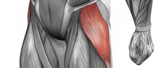
Exercises for the muscles of the buttocks
We're done with the "gunpowder", let's move on to the "berries". anatomy of the buttocks:
Exercises for the muscles of the buttocks:
List of exercises for the gluteal muscles:
- Lunges with a barbell on the shoulders
- Lunges with dumbbells
- Swing your leg back from a low block
- Swing your leg back with a machine lever
- Swing your leg back on the floor
- 'Bridge' lying down
- Swing your leg to the side from the lower block
- Swing your leg to the side with the machine lever
- Swing your leg to the side while lying on your side
- Breeding legs on the simulator
Here it is worth paying attention to the instability of the knee joint and the individual mobility of the hips.