A pinched nerve in the thoracic region is a rare occurrence, since it is the most immobile part of the spinal column and does not bear a large load. Pinching can occur as a result of a sudden movement, or it can be a consequence of complications of various pathologies (for example, osteochondrosis of the thoracic region). This disease significantly reduces the quality of life, so it is necessary to take timely treatment seriously.
Description of the pathology
Pinching of the thoracic nerve in the spine is accompanied by severe pain. Its nature depends on the type of pinched nerve.
They come in 3 types: one is responsible for motor functions, the other for sensitivity, and the third for autonomic function.
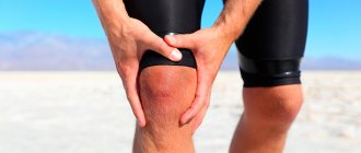
The main cause of a pinched nerve in the chest area is the progression of intercostal neuralgia. Any sudden movement or lifting of heavy weights can provoke pinching in the spine. Also, the disease can occur in the stage of exacerbation of the spine.
Dystrophic changes cause displacement of the vertebrae and compression of the nerves. Sometimes it is provoked by joint diseases (arthritis, arthrosis). Pinching in the thoracic spine can lead to a number of negative consequences, so it is important to consult a doctor promptly.
Main risk factors
Surgical intervention does not exclude complications, and serious ones at that. Especially if mistakes were made during the intra- and/or postoperative period. Even small errors during surgery or during rehabilitation increase the likelihood of unsatisfactory hip arthroplasty.
- advanced age of a person;
- severe concomitant disease, for example, diabetes mellitus, arthritis of rheumatoid etiology, psoriasis, lupus erythematosus;
- any previous surgical intervention on the “native” joint aimed at treating dysplasia, femoral fractures, coxarthrosis deformities (osteosynthesis, osteotomy, etc.);
- re-endoprosthetics, that is, repeated replacement of the hip joint;
- local inflammation and purulent foci in the patient's history.
It should be noted that after hip joint replacement, elderly people are more susceptible to complications, and especially those over 60. In addition to the underlying disease, elderly patients have concomitant pathologies that can complicate the course of rehabilitation, for example, reduce resistance to infection.
It is more difficult for older people to recover, but this can be done successfully.
Installation of a prosthesis requires removal of the ligaments that stabilize the joint. Therefore, after the operation, only the muscles will be responsible for keeping it in the correct position. If they do not function sufficiently, for example, due to underdevelopment and lack of training, and the patient allows himself dangerous sudden movements, this often leads to dislocation of the joint.
- normal bending of the leg, for example, while sitting or raising the knee;
- extension - during the swing when hitting the ball;
- crossing the legs inward, or adducting;
- moving the leg to the side;
- rotation inward or outward (adduction and expansion of the feet with straight legs).
Symptoms
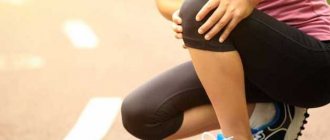
When a nerve is pinched in the thoracic region, symptoms usually appear immediately. Due to pinching of the nerve, a person experiences severe pain in the chest area. They can be of different nature (sharp, encircling, pulsating) and intensity. Often this pain is confused with cardiovascular diseases, since a person begins to have breathing problems and severe shortness of breath.
When pinched, disruptions occur in the transmission of nerve impulses from organs to the nervous system. As a result, a person may experience tingling and numbness in the upper limbs and back area. Weakness in the hands is also noted. In some cases, temporary paralysis occurs (it goes away after treatment).
Pinching negatively affects the gastrointestinal tract, causing severe imbalance. The symptoms resemble colitis and ulcers: cramps and severe pain in the abdominal area.
Types of joint fixation
In orthopedics and traumatology, 3 methods of fixation have been developed, these are:
- cement;
- cementless;
- combined.
With cement bonding, your new joint will be “seated” on medical-grade acrylate-based cement. The mixed solution hardens very quickly, in 10 minutes, forming a dense monolith that is extremely durable.
The cement solution is injected into the channel of the tubular element and the recess of the iliac bone, and then the structural units included in the endoprosthesis are “planted” onto it. This technique is best suited for older people and patients with osteoporosis. The fixed surfaces of such implants are glossy.
Acetabular components of the hip implant, pink surface made of ceramic, white surface made of polyethylene.
Cementless fixation is the most common technique. Its principle is to hammer parts with a rough surface into prepared bones. Naturally, a strong connection with the bones will not occur immediately; the implant must be well overgrown with connective tissue structures, which will take up to 3 months.
Zimmer endoprosthesis.
Combined prosthetics is a combination of two considered technologies simultaneously during one operation. This version of the technology is intended for those who have different bone densities in the hip and pelvis.
Osteoporosis, as you can see, makes the bone looser.
It is the condition of the bone that determines what type of fixation will be used.
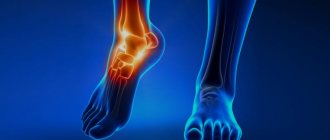
How to diagnose a pinched nerve in the thoracic spine
If characteristic signs of the disease appear, you need to seek help from a specialist. The problem is treated by a neurologist. At the initial appointment, he will collect a complete medical history and conduct a visual and tactile examination of the spine to determine the degree of sensitivity of specific areas of the body.
The next stage is an instrumental examination. In order to assess the condition of the thoracic region, identify bone deformities or an infectious process, X-ray examination, computed tomography and magnetic resonance imaging are prescribed. Based on the research results, the doctor makes a diagnosis. In some cases, differential and laboratory diagnostics (general blood and urine tests) may be necessary.
Hospitalization
All necessary information regarding preoperative preoperative measures is provided by the attending doctor. Also at this stage, the surgeon decides on the choice of endoprosthesis and plans every step, and the anesthesiologist selects the appropriate type of anesthesia based on the patient’s health indicators.
At the clinic, the admitted patient is carefully examined by various specialists:
- examination by a primary specialist (orthopedist-traumatologist);
- X-ray, MRI, ultrasound of the hip joint, if necessary, endoscopic examination of the problem area;
- consultation with highly specialized doctors (therapist, cardiologist, immunologist, anesthesiologist, dentist, gastroenterologist, etc.);
- general and biochemical blood test;
- coagulogram (determines the functioning of the hemostasis system, in particular the mechanism and time of blood clotting);
- test for blood group and Rh factor;
- clinical urine analysis;
- fluorography;
- electrocardiogram.
First aid
In most cases, a pinched nerve occurs suddenly and is accompanied by severe pain. First of all, you need to calm down, breathe calmly and lie down on a hard surface so that the least pain is felt. In order to eliminate the pain, you need to take a painkiller. If the pinching caused strong emotional stress, then you can take a sedative.
If the pain from the pills does not go away, but begins to intensify, it is advisable to call emergency help. When she arrives, it is important to notify the doctor about all previously taken medications so that further treatment is effective.
Preventive measures
Complications after hip replacement are much easier to prevent than to then undergo labor-intensive and lengthy treatment to get rid of them. Unsatisfactory development of the situation can nullify all the efforts of the surgeon.
Infections are treated with antibiotics, which in itself is quite harmful to the body.
At the preoperative stage, diagnostics are performed for infections in the body, diseases of internal organs, allergies, etc. If inflammatory and infectious processes are detected, chronic diseases in the stage of decompensation, surgical measures will not begin until the identified foci of infections are cured, venous-vascular problems will not be reduced to an acceptable level, and other ailments will not be brought into a state of stable remission.
Currently, almost all implants are made from hypoallergenic materials.
If there is a predisposition to allergic reactions, this fact is examined and taken into account, since the choice of medications, endoprosthesis materials and type of anesthesia depends on it. The entire surgical process and further rehabilitation are based on assessing the health status of internal organs and systems, age criteria and weight.
To minimize the risks of complications after hip joint replacement, prophylaxis is carried out before and during the procedure, after surgery, including the long-term period. Comprehensive preventative approach:
- drug elimination of the infectious source, full compensation of chronic ailments;
- prescribing certain doses of low molecular weight heparins 12 hours in advance to prevent thrombotic events; antithrombotic therapy continues for some time after surgery;
- the use of broad-spectrum antibiotics active against a wide group of pathogens a couple of hours before the upcoming hip replacement and for several days;
- technically impeccable surgical intervention, with minimal trauma, avoiding significant blood loss and the appearance of hematomas;
- selection of an ideal prosthetic structure that completely coincides with the anatomical parameters of the real bone connection, including its correct fixation at the correct orientation angle, which in the future guarantees the stability of the implant, its integrity and excellent functionality;
- early activation of the patient in order to prevent congestion in the leg, muscle atrophy and contractures, inclusion of exercise therapy and physiotherapy procedures (electromyostimulation, magnetic therapy, etc.), breathing exercises from the first day, as well as high-quality care of the surgical wound;
- informing the patient about all possible complications, permitted and unacceptable types of physical activity, precautions and the need to regularly perform physical therapy exercises.
Communication between the patient and the medical staff plays a huge role in successful treatment. This is what is called service, because when the patient is fully instructed, he better perceives the processes occurring in his body.
The patient must realize that the outcome of the operation and the success of the recovery depend not only on the degree of professionalism of the doctors, but also on himself. After hip replacement surgery, it is possible to avoid unwanted complications, but only if you strictly follow the recommendations of specialists.
Treatment of a pinched nerve in the thoracic spine
As a rule, treatment of pinched spine is carried out using conservative methods. The choice of method depends entirely on the reason that caused the pinching. At the initial stage, medication and physiotherapeutic methods are used. Let's take a closer look at the specifics of treating spinal impingement.
Drug therapy
In most cases, drug treatment is used at the beginning of therapy. It includes taking the following:
- muscle relaxants. They are prescribed to eliminate pain and muscle spasms,
- anti-inflammatory drugs. They eliminate the inflammatory process from the affected area,
- sedatives. Such drugs have a positive effect on the nervous system,
- B vitamins and microelements. They affect the regeneration of damaged nerve fibers. For greater effectiveness, injections are prescribed rather than tablets.
Physiotherapeutic methods
After the pain has subsided, the doctor may prescribe physiotherapeutic treatment. It cannot be performed in the acute stage of the disease. Most often everyone uses:
- electrophoresis,
- peloidotherapy,
- ultraviolet irradiation,
- magnetic therapy,
- acupuncture.
All of the above procedures are effective, they allow you to achieve nerve release and normalize blood circulation, which significantly improves metabolic processes.
The main contraindications are high body temperature, malignant neoplasms and some pathologies of the cardiac system. Also, nerve release is achieved through manual therapy and massage courses. These procedures can only be performed if there is no pain.
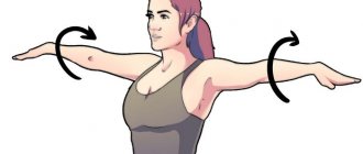
In the event that the disease is caused by dystrophic-degenerative changes in the spine, courses of therapeutic exercises are prescribed. They are aimed at restoring the correct physiological position of the thoracic region. With the help of special exercises, the muscles of the spinal column are significantly strengthened, thereby reducing pressure on the vertebrae.
The attending physician will select the most appropriate exercises depending on the age, physical condition of the patient and the stage of the disease. If severe discomfort or other negative symptoms occur, you should stop exercising and report these signs to your doctor so that he can select other exercises.
Surgical intervention
Surgical intervention is resorted to when it was not possible to achieve results with conservative treatment. For example, if diagnostics reveals a hernia, in 90% of cases surgery is prescribed. In addition, benign and malignant neoplasms, advanced stage osteochondrosis and serious injuries are subject to surgical intervention.
There are different methods of surgical intervention, the most suitable method is chosen depending on the detected pathology. Surgeries can be radical (complete removal of a spinal tumor) or minimally invasive. In order to remove a protruding portion of the disc, they most often resort to discectomy.
Folk remedies
Traditional medicine is ineffective as the only method of treatment; it is more advisable to use them along with the methods described above. The most popular are various infusions, decoctions and healing baths. Let's look at the recipes for the most effective remedies:
- Infusion (herbal collection). To prepare the infusion, take thyme, rose hips, linden and hawthorn in a ratio of 5:4:2:2. Grind all ingredients (2 tablespoons) and pour 400 ml of boiling water. Leave to infuse for 2-3 hours and consume 200 ml 2 times a day.
- Spruce bathtub. Conifer bark (500 g) is added to 3 liters of water and placed on low heat. The product should be boiled for about 20 minutes, then filtered and added to the bath.
- Infusion based on lingonberries. To prepare the product, take 1 tablespoon of plant leaves and pour 2 liters of boiling water. After 2-3 hours, consume 100 ml 2-3 times a day.
- A decoction of orange peels and lemon balm. Take the indicated ingredients in equal proportions (2 teaspoons each) and pour in 500 ml of hot water. Leave for 20 minutes, then filter and add 2 teaspoons of valerian tincture. Drink 250 ml per day.
- Wraps. Take horseradish leaves and apply to the damaged area. Wrap the top with cling film and cover with a warm cloth. Keep this wrap for no more than 4-6 hours.
As a rule, natural ingredients are harmless to health, but may cause individual intolerance.
In order not to harm the body, it is recommended to consult with your doctor before using folk remedies.
Minimally invasive method
Symptoms of complications after hip replacement will be presented below in the table for better understanding. A quick visit to a doctor at the first suspicious signs will help to avoid the progression of adverse events, and in some situations, to save the implant without revision surgery.
Negative excess occurs in the first year after prosthetics. This is the most common pathological condition in which the femoral component is displaced in relation to the acetabular element, resulting in separation of the head and cup of the endoprosthesis.
Dislocation of the femoral component on x-ray.
The risk group includes people with hip fractures, dysplasia, neuromuscular pathologies, obesity, joint hypermobility, Ehlers syndrome, and patients over 60 years of age. Individuals who have undergone surgery on a natural hip joint in the past are also particularly vulnerable to dislocation.
The dislocation requires non-surgical reduction or open repair. If treated in a timely manner, it is possible to adjust the endoprosthetic head using a closed method under anesthesia. If the problem continues, the doctor may prescribe a repeat operation to reinstall the endoprosthesis.
The second most common phenomenon, characterized by the activation of severe purulent-inflammatory processes in the area of the installed implant. Infectious antigens are introduced intraoperatively through insufficiently sterile surgical instruments (rarely) or after intervention they move through the bloodstream from any problematic organ that has a pathogenic microbial environment (often).
Discharge from a surgical wound is a bad sign.
A purulent focus has a detrimental effect on the strength of fixation of the endoprosthesis, causing its loosening and instability. Pyogenic microflora is difficult to treat and, as a rule, requires removal of the implant and re-installation after a long time.
The arrows indicate areas of infectious inflammation, this is exactly what they look like on an x-ray.
PE is a critical blockage of the branches or main trunk of the pulmonary artery by a detached thrombus, which formed after implantation in the deep veins of the lower limb due to low blood circulation resulting from limited mobility of the leg.
This complication is dealt with quite successfully at this stage of medical development.
Blocking the lumen of the lungs is dangerously fatal, so the patient is immediately hospitalized in the intensive care unit, where, taking into account the severity of the thrombotic syndrome: administration of thrombolytics and drugs that reduce blood clotting, NMS and mechanical ventilation, embolectomy, etc.
It is technically much easier to operate if the surgical wound is large. Therefore, orthopedists and traumatologists argue that the classical procedure on large joints is still an undeniable priority.
Many clinics practice a minimally invasive approach, using an incision almost 2 times smaller than with traditional intervention. However, advanced surgeons with sufficient experience increasingly agree that it is better not to take unnecessary risks, but to use proven, traditionally established tactics, the standard of prosthetics - to create a neat incision from a direct lateral approach, approximately 13 cm long.
According to the minimally invasive technique, the incision is approximately 7 cm. The most commonly used approaches, which provide a less traumatic approach from a physiological point of view, are postero- and anterolateral. The advantages of the minimally invasive type are the preservation of the abductor muscles, the compactness of the surgical wound, and the accelerated rate of rehabilitation recovery.
Clinical studies show that intraoperative blood loss with gentle intervention is insignificant, pain syndrome in the postoperative phase is less pronounced compared to the standard procedure.
Visual comparison of incision sizes using the classical (right) and minimally invasive (left) techniques.
Complications
If treatment is not provided in a timely manner, a person may experience various complications. Pinching in the spine can lead to the following complications: impaired motor function of the limbs, chronic back pain, bulging discs and paralysis.
If you carry out high-quality treatment and follow all the doctor’s recommendations (for example, do physical therapy exercises), then pinching in the spine will not lead to negative consequences.
Types of endoprostheses
- Deep armchairs. In them, the body position turns out to be unfavorable for the joint, the load is far from natural and complications are possible.
- Slippery surfaces. The likelihood of injury increases significantly.
- Deep squats. In the absence of stabilizing ligaments, the muscles may not be able to cope with the increased load.
- Sitting on a chair diagonally. Only natural positions are permissible - diagonally the load on the body is distributed unevenly, which is very dangerous.
- Sitting with crossed legs. Another test for the endoprosthesis.
- Sharp turns of the body. Risk of mechanical injuries.
- Lifting weights. Now we will have to forget about this forever.
- Support on the sore leg. After endoprosthetics, you will have to ensure that your body weight is distributed evenly. Standing on your sore leg is very dangerous.