Fast and slow muscle fibers
Fast-twitch muscle fibers (glycolytic) are fast-twitch fibers that are strong but fatigue-resistant. For ease of perception, we will shorten their name to the officially accepted abbreviation - GMV .
Slow-twitch muscle fibers (oxidative) are slow-twitch fibers; on the contrary, they are characterized by low strength and low fatigue. For ease of perception, we will shorten their name to the officially accepted abbreviation - OMV .
In our body, everything is thought out to the smallest detail, and muscles are no exception. Depending on the duration and intensity of the load, certain muscle fibers are involved, and their ratio directly affects our athletic achievements. That's why the information below is essential to building every athlete's training program!
Similarities
Both red and white fibers belong to muscle tissue and are part of the same muscles. Both types of fibers are tools for achieving specific goals . Switching between slow and fast mechanisms occurs automatically, as needed, and completely unnoticed by us.
For successful work, both red and white fibers require a sufficient amount of energy; without its replenishment, the muscles will not work. In both cases, energy synthesis occurs in mitochondria, which are located inside muscle cells. One such cell may contain several thousand mitochondria. These energy synthesizers are lined up in chains along thin threads - myofibrils, which provide muscle contraction processes.
GMV vs OMV
Most likely, you have already heard that the fibers that make up our muscles are of two types: fast-twitch (FMV) and slow-twitch (SMV). More precisely, there is also a third, intermediate type - transition fibers.
The type of fiber is determined by the number of nerve impulses sent to the fiber. The more impulses there are, the correspondingly higher the activity of adesine triphosphatase, as well as the higher the speed of fiber contraction.
Adesine triphosphatase is a special enzyme class of hydrolases that accelerates the process of cleavage of H3PO4 from adenosine triphosphate molecules, which results in the release of energy used for muscle contraction.
GMW (white)
So why are they "white"? It's all about the capillaries they contain, which are much smaller than in OMV, hence the differences in color. In terms of its structure, GMV is, as a rule, several times thicker than OMV. Their response to signals coming from the brain is instantaneous, and the rate of contraction is at least twice as high as that of oxidative ones. Glycolytic fibers receive energy from rapidly absorbed ATP, creatine phosphates and glycogen. It is necessary to understand that these energy sources dry up in just 30-60 seconds. In the process of obtaining energy by fast fibers, oxygen is not involved, due to which energy is released almost instantly, but its reserves are very limited. Based on this, we can conclude that white muscle fibers are suitable for high-intensity, but short-term loads. However, their energy is not enough to perform numerous repetitions and long, monotonous movements.
OMV (red)
They are the exact opposite of glycolytic in their structure and function, and are literally created for light and prolonged loads. They are able to accumulate, store energy, and then gradually expend it, thanks to mitochondria and myoglobin. So, if your muscles are dominated by RMF, you may well be a long-distance runner, aerobic sports are also suitable for you.
Unfortunately, OMVs have much less potential for growth in their volumes and quantities than glycolytic ones. So the increase in our muscle mass is mainly due to GMV.
The ratio of OMV and HMV in our body is predetermined by genetics and we cannot change it. The vast majority of us are dominated by oxidative fibers; in every fourth person, on the contrary, the percentage of glycolytic fibers is slightly higher than red fibers. And only in some athletes the predominance of some muscle fibers over others reaches 85% - they are the ones who have the highest chances of achieving the greatest results in sports.
Muscles. general review
1. What is the active part of the musculoskeletal system?
The active part of the musculoskeletal system includes muscles.
2. Remember what types of muscle tissue are found in the human body. Which of them are the muscles of skeletal muscles formed?
There are 3 types of muscle tissue in the human body: striated, cardiac, smooth. Skeletal muscle is formed by striated muscle tissue.
3. How are muscles attached to bones?
Muscles are attached to bones by inextensible tendons that fuse with the periosteum. Muscles can be attached either only on one side to the bone and the other to the skin, or intertwined with another muscle, or attached to the bones at both ends. In this case, the muscles are attached at one end above and at the other below the joint. With this type of attachment, muscle contraction moves the bones in the joints.
4. Explain the mechanism of contraction of striated fibers. Why were they called that? How is the process of contraction and relaxation of muscle fibers regulated?
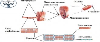
Each muscle fiber is a multinucleated cylindrical cell. The diameter of these cells ranges from 5 to 100 microns, the length reaches 10-12 cm. The general structure of a muscle cell is the same as that of other animal cells, only all structures are shifted towards the cell membranes, and in the center there are numerous thin contractile filaments - myofibrils. Myofibrils are formed by two types of contractile proteins - actin (thinner and lighter) and myosin (thicker and darker). These proteins are arranged in myofibrils in an orderly manner so that myosin molecules fit into the spaces between actin molecules. Therefore, dark and light areas alternate in the myofibril. Hence the name of skeletal muscles - striated. At the moment when an electrical signal comes from the nervous system to the muscle along the nerve fiber, the electrical potential changes on the cell membrane, which causes the release of calcium molecules from the sarcoplasmic reticulum (calcium depot in the muscle cell). The calcium molecule interacts with actin, which causes the actin processes to shift from the blocking position, which makes it possible for actin molecules to interact with myosin molecules. Myosin filaments go deeper into the spaces between actin molecules - the muscle contracts and thickens. The movement of myosin causes the activation of ATPase located on it and the release of ATP molecules, due to which the bond between actin and myosin is broken. In parallel, calcium reenters the sarcoplasmic reticulum and the muscle relaxes.
5. What is the difference between red and white muscle fibers?
They differ in the composition and number of myofibrils, which determines the characteristics of their contraction. The so-called white muscle fibers contain less of the protein myoglobin, which provides color, contract quickly, but quickly and fatigue; red fibers contain a large amount of myoglobin, contract more slowly, but can remain in a contracted state for a long time. Depending on the function of the muscles, certain types of fibers predominate in them. White fibers play a major role in gaining mass in bodybuilders, and also provide runners and sprint swimmers with maximum speed. Red fibers are perfect for low-intensity and long-term work, such as walking and light running, distance swimming, aerobics, etc.
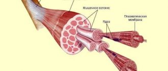
6. How is skeletal muscle structured? What structures besides muscle fibers does it contain?
The muscle fibers of striated muscle tissue are collected in bundles, which contain 10-50 fibers. These bundles are surrounded by connective tissue (fascia). The muscle itself is also surrounded by fascia. The mass of muscle tissue in skeletal muscle is about 90%. Muscles also include blood vessels that carry nutrients to muscle fibers and metabolic products from them, and nerve fibers through which a signal for muscle contraction comes from the brain.
7. What groups can skeletal muscles be divided into?
Depending on their location, muscles can be divided into the following large groups: muscles of the head and neck, muscles of the trunk and muscles of the limbs. In turn, the head muscles are divided into chewing and facial muscles. The muscles of the trunk include the muscles of the chest walls, abdomen and back.
8. What are the features of attachment of facial muscles?
Facial muscles differ from all skeletal muscles in that they are attached to the bones of the skull at one end and to the skin at the other. Therefore, when they contract, the shape and depth of the skin folds change. Facial muscles are mainly located around natural openings - oral, eye, ear, nasal and are anatomically independent from each other.
In some people, on one or both sides, the levator anguli oris muscle is not only attached to the skin at one of its ends, but also with part of the fibers between the two ends. With this passage, when the muscle contracts, it not only lifts the corner of the mouth, but also pulls along a section of the skin of the cheek. Then, when a person smiles, “dimples” appear on his cheeks.
9. Why are there large muscles on the shoulder, and many small muscles on the forearm?
The shoulder muscles move the entire arm: flexion and extension, rotation at the elbow joint. Movements in this joint do not require great precision, but, since they lift the mass of the entire arm, they require great strength, which is why these muscles are large and few in number. The muscles of the forearm are responsible for the movements of the hand and fingers. These are “small” and precise movements, therefore the muscles are small, numerous, with a large number of tendons.
10. Name the longest muscle in our body.
The longest muscle is the sartorius muscle, located on the thigh. It was so named because it was also “pumped up” by tailors as they worked at the foot-operated sewing machine.
11. Describe the functions of the muscles indicated in the figure on p. 120 textbook.
Deltoid muscle: takes part in flexion and extension of the shoulder, abduction of the arm to the side.
Biceps brachii: flexes the arm at the elbow and rotates the palm toward the body (supination) when the arm is bent.
Triceps brachii muscle: due to the long head, the arm moves backward and brings the arm to the body; the entire muscle takes part in the extension of the forearm.
Extensor muscles of the hand and fingers: the posterior group of muscles of the forearm, is involved in the extension of the wrist joint and fingers (along the phalanges).
Flexor muscles of the hand and fingers: muscles are antagonists of the posterior group of muscles of the forearm. Participate in flexion of the wrist joint and fingers (along the phalanges).
The pectoralis major muscle: leads the shoulder to the body and turns the arm inward, that is, it pronates the arm, and is an auxiliary muscle of inspiration.
Serratus anterior: pulls the scapula away from the spinal column; together with the rhomboid muscle, it fixes the scapula to the surface of the chest. With a stationary girdle of the upper limb, the serratus anterior muscle is also an auxiliary muscle of inspiration.
Trapezius muscle: brings the scapula closer to the spinal column, contracting with all bundles, raises the scapula, contracting with the upper bundles, and lowers, contracting with the lower ones.
Latissimus dorsi: adducts the shoulder to the torso and pulls the upper limb back toward the midline, rotating it inward. If the upper limb is fixed, it brings the torso closer to it and can expand the chest, serving as an auxiliary respiratory muscle.
Abdominal muscles: participate in building the anterior abdominal wall, flexing the torso, and are additional respiratory muscles.
Back extensor muscles: keep the spine straight.
Gluteus maximus: extends the flexed hip, participates in external rotation of the hip, abducts the hip.
Biceps femoris muscle: flexes and externally rotates the lower leg, extends the thigh at the hip joint.
Sartorial muscle: abducts, externally rotates and flexes the thigh; bends the lower leg at the knee joint.
Quadriceps muscle: extends the tibia at the knee joint.
Tibialis anterior: Involved in dorsiflexion of the foot.
Calf muscle: provides plantar flexion of the foot, helps flex the lower leg at the knee joint, raises the heel when walking.
Muscle fiber training
The main goal of bodybuilders is to increase muscle mass, which mainly depends on the growth of muscle mass.
Glycolytic fibers
To increase their volume, intense short-term loads are used using large weights (60-80% of the repeated maximum) and with constant alternation of muscle groups. The cross-section of fibers increases, as well as energy reserves in the muscles, resulting in muscle hypertrophy.
The duration of one approach is less than a minute. Rest time between sets is 2-4 minutes. Average training frequency – three strength training days a week is quite enough. Exercises are performed at an average pace, neither fast nor slow, with full amplitude; individual phases of the exercises are not highlighted.
Oxidative fibers
The exercises are performed with a light weight of 30-50% of the weight with which you can perform the exercise with only one repetition. An average of 15 to 30 repetitions are performed per set. Approaches 5-8, more possible. It is necessary to perform exercises at a slow or medium pace, without highlighting certain phases of movement. The amplitude of the exercises is full.
Muscle fibers: how to determine your type
Typically, a person has approximately 40% slow fibers and 60% fast fibers. Their exact number is determined genetically. Analyze your physique and perception of stress. As a rule, people who are naturally “wiry”, short in stature, with thin bones, who can easily walk, jog, ride a bike and other long-term activities, have a slightly higher percentage of slow and intermediate fibers.
And those who have wide bones, muscles easily grow even from small loads, but also the fat layer is added literally from one glance at cakes or pasta, are often “carriers” of some excess of fast-twitch fibers. If you know a person who, without really training, suddenly amazes everyone with his strength, you have a large number of fast, non-oxidative fibers. You can find tests online that offer to determine your predominant muscle fiber type. For example, doing an exercise with a weight of 80% of the maximum. If you completed less than 8 repetitions, your fast-twitch fibers predominate. More - slow.
In fact, this test is very conditional and speaks more about training in this particular exercise.
Fibers illustrated
In order to fully understand what GMV and OMV are and what they look like, there is nothing better than seeing them with your own eyes. And it's very easy to do. Do you eat chicken? The fact is that it is chicken meat that best reflects the location of glycolytic and oxidative fibers in the poultry body. Surely many of you have noticed that chicken meat in the area of the breast and wings is white, and it contains practically no fat, while the meat of chicken legs and thighs has a dark red color and a higher fat content. The thing is that a chicken, like most other poultry, spends almost all its time standing, which means that the muscles of its legs are subject to constant static load (i.e., oxidative fibers are involved). At the same time, the wings are used extremely rarely and only for quick, energetic flapping, which characterizes the work of glycolytic fibers.
White fibers
This color of the fibers is due to too little coloring pigment in them - myoglobin and small capillaries. According to energy, these fibers are divided into two subtypes: 2A and 2B . The first subtype 2A receives energy both as a result of anaerobic glycolysis (without oxygen) and aerobic (with the participation of oxygen). But the energy of subtype 2B is aimed only at anaerobic glycolysis.
White muscles come into play when there is a lot to be done in a short period of time . They can contract at high speed, while giving power and strength. White muscles are indispensable in sports, for example, when in a split second you need to make a breakthrough, overcome yourself and set a world record.
But such a speed limit cannot last long, and there are a number of reasons for this:
- Energy reserves are not endless; during intensive work they do not last long.
- To restore wasted energy, the body needs time, at least 2-5 minutes - during this time the required amount of creatine phosphate ATP molecules is usually produced.
- With each repetition of an intense contraction and the receipt of a new portion of energy, lactic acid is produced during the reaction process, which causes pain and loss of strength.
Both subtypes: 2A and 2B - harmoniously complement each other. White fibers, classified as subtype 2A, contract quickly and have great strength. 2B – work even faster, achieving enormous power and strength. The source of energy for white fibers is glycogen, obtained from the breakdown of glucose, and creatine phosphate, which enters the body along with protein foods.
Muscle contraction speed
The speed of muscle contraction is the time it takes for muscle tissue to respond to an irritating impulse from the nervous system.
Skeletal muscles are riddled with nerve endings. Each muscle thread is covered by a motor plaque. Through these connections, the brain sends commands to the muscles - nerve impulses. The more impulses, the higher the rate of muscle fiber contraction.
Mitochondria and myofibrils in muscles
Let's look at the structure of muscle fiber. Its cytoplasm (sarcoplasm) contains a large number of mitochondria. They play the role of power plants in which metabolism occurs and energy-rich substances accumulate, as well as those needed to meet energy needs. Any muscle cell contains several thousand mitochondria. They occupy approximately 30-35% of its total mass.
The structure of muscle fiber is such that a chain of mitochondria is lined up along the myofibrils. These are thin threads that provide contraction and relaxation of our muscles. Typically, one cell contains several dozen myofibrils, and the length of each can reach several centimeters. If you add up the mass of all myofibrils that make up a muscle cell, then its percentage of the total mass will be about 50%. The thickness of the fiber thus depends primarily on the number of myofibrils contained in it, as well as on their cross-sectional structure. In turn, myofibrils consist of a large number of tiny sarcomeres.
Cross-striped fibers are characteristic of muscle tissue of both women and men. However, their structure differs slightly depending on gender. Based on the results of a biopsy of muscle tissue, it was concluded that the percentage of myofibrils in the muscle fibers of women is lower than in men. This applies even to high-level athletes.
By the way, muscle mass itself is distributed unequally throughout the body in women and men. The vast majority of it in women is located in the lower part of the body. At the top, the muscle volumes are small, and they themselves are small and often completely untrained.
Features of red fibers
Slow-twitch muscle fibers have many mitochondria. They carry out the oxidation process, which is necessary to produce energy. The red fibers are surrounded by a large network of capillaries. They are needed to deliver large volumes of oxygen along with blood.
Slow muscle fibers are well adapted to the implementation of the aerobic energy generation system. The strength of their contractions is relatively small. The rate at which they consume energy is sufficient to make do with aerobic metabolism alone. Red fibers are perfect for low-intensity and long-term work, such as walking and light running, distance swimming, aerobics, etc.
The contraction of muscle fiber allows for movements that do not require much effort. Thanks to it, the posture is also supported. These striated fibers are characteristic of muscle tissues that are activated under loads ranging from 20 to 25% of the maximum possible force. They are characterized by excellent endurance. However, red fibers do not work when performing sprints, lifting heavy weights, etc., since these types of loads require a fairly rapid expenditure and production of energy. White fibers are intended for this, which we will talk about now.
Fast glycolytic fibers of muscle tissue
The first type is fast glycolytic fibers. They use the process of glycolysis to produce energy. In other words, they are able to use only the anaerobic energy production system, which promotes the formation of lactic acid (lactate). Accordingly, these fibers do not produce energy with the participation of oxygen, that is, aerobically. Fast glycolytic fibers are characterized by maximum contraction speed and strength. They play a major role in bulking for bodybuilders and also provide maximum speed to runners and sprint swimmers.
White and red fibers are the main
Despite the fact that these two types of fibers are diametrically opposed, they always work in conjunction, replacing each other.
For example, you are training in the gym and started lifting a heavy barbell (no matter how), which in your training you cannot lift more than 2-3 times in one approach (set). In this case, the blow is taken by the white fibers, which are responsible for physical work in an explosive style. That is, you can lift heavy weights, but not for long.
Second example: You took a significant amount of weight off the barbell and started lifting it again. Now you feel ready to do up to 20 repetitions, and you think you can do even more. In this case, red muscle fibers work, allowing the body to perform long-term work. After all, doing 3 repetitions and 20 is a big difference in time.
Third example. You've loaded the barbell with about three-quarters of the weight you can lift at one time (no more) - that's 75%. In this exercise you can do 10-12 repetitions. I will say that in the first lifts of the weight, white fibers actively work, at the end of the exercise the white fibers weaken significantly (we feel tired, but we can continue working), and the red ones are just beginning their work. This is what I was talking about - they replace each other.
Below I will describe in more detail why things are this way.
If these two types are given specific characteristics, then white is strength, red is endurance.
If you think, like I did at one time, that there are only two types of fibers, then you are mistaken. There is also an intermediate type that contains the characteristics of the other two types. He can be both strong and resilient at the same time.
By the way, did you know that the number of certain fibers is not constant in our body? I would like to bring to your attention, friends, that a certain type of training (if you train systematically for a long time) can change the ratio of fibers: today red fibers predominate, and in six months white fibers predominate. This means that you were hardy, but after training for six months, you became strong.
Features of white fibers
White fibers are characterized by a larger diameter relative to red ones. In addition, they contain much more glycogen and myofibrils, but they have fewer mitochondria. This type of muscle fiber cell also contains creatine phosphate (CP). It is required at the initial stage of high-intensity work.
White fibers are best suited for performing powerful, fast, but short-term efforts, since they have low endurance. Fast fibers, compared to slow fibers, are able to contract 2 times faster, and also develop force 10 times greater. It is thanks to them that a person develops maximum speed and strength. If the work requires 25-30% of maximum effort and above, this means that it is white fibers that take part in it. They are divided according to the method of obtaining energy into the following 2 types.