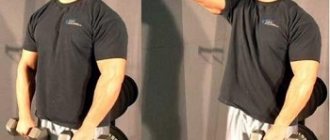
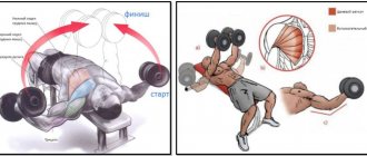
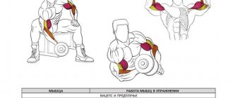
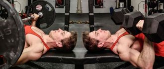
This exercise perfectly outlines the biceps and gives it a sporty and sculpted shape. The essence of supination is the additional rotation of the hands when lifting dumbbells for biceps, which cannot be done with a barbell. This technique maximally engages all the fibers of the biceps, and with different variations of the exercise, you can additionally engage the anterior deltoid and upper part of the pectoral muscle.
Pronation and supination anatomy. Supination, pronation and VPN exercises
Supination, pronation and VPN exercises
If you carefully study the anatomy of muscles and especially the functions they perform, you can learn a lot of interesting and practically useful things.
In this article, we'll delve deeper into the muscles that control the shoulder joint. There is no doubt that the shoulder joints are extremely important in everyday life. In essence, this is the place from which hands grow, with all the ensuing consequences
When doing fitness, remembering the condition of your shoulder joints is one of the signs of caring about your health and a manifestation of an elementary training culture.
What is supination and pronation?
To clearly understand what we are talking about here, you should understand these terms - supination and pronation. They are often found in books and articles on training.
Supination is an outward rotational movement of a limb or part of it. For example, if there is supination of the hand, bent at the elbow at an angle of 90 degrees, then the hand turns from a palm down position to a palm up position. In the figure this is indicated by number 1.
And it is not so important how the arm is bent at the elbow. Rotation in the direction indicated by arrow 1 is always supination.
Pronation is the opposite of supination, a rotational movement that occurs inward. If you hold a handful of coins in your palm and then decide to pour them out, you need pronation.
The use of the terms pronation and supination in relation to the hand is more or less clear. They are especially popular on bodybuilding forums when it comes to training the biceps and forearms.
What about shoulder pronation and supination?
We hear about this much less often. And completely in vain. In this picture, the athlete performs supination of the shoulder joints.
And on this pronation.
What is VPN?
VPN stands for shoulder external rotation. That is, shoulder supination.VPN exercises are exercises that develop the muscles that rotate the shoulder outward (shoulder supinators).
Why is this necessary?
At the beginning of the article, I drew your attention to the study of muscles and their functions. We read about the muscles that control the shoulder joints:
Shoulder pronation: 1) subscapularis, 2) pectoralis major, 3) anterior deltoid, 4) latissimus dorsi, 5) teres major. Shoulder supination: 1) infraspinatus, 2) teres minor, 3) posterior deltoid. muscles
If you look at the drawing carefully, a lot will become clear. A – front view, B – rear view.
Note! The pronators of the humerus are the two largest muscles in the body - the pectoralis major and the latissimus dorsi! And also the anterior deltoid. During training, everyone does nothing but develop these muscles in every possible way. What happens?
It turns out the following. From strength training, muscles shorten and become noticeably stronger and increase in volume. Have you noticed how funny the arms of massive bodybuilders diverge to the sides? One of the reasons for this is the penetrating effect of the pumped pectoral and latissimus muscles.
The shoulder joint gradually more and more comes into a state of slight, but still noticeable pronation. This is not a completely natural position, and the shoulder joint should bear the main load in a neutral position, and not in a supinated or pronated position.
If we constantly do nothing but pump up the pronators of the shoulders, we create all the prerequisites for distorting the normal geometry of our own shoulder joints. From here it’s not far to injury. Especially when performing bench presses, bent-over rows, and pull-ups. Once! And a sharp pain in the shoulder!
What to do?
1. Include in your training exercises that develop shoulder supinators. This is VPN exercise. They develop mainly the muscles lying on the back of the shoulder blades - supraspinatus, infraspinatus, teres minor.
I will tell and demonstrate them in one of the upcoming articles.
2. Practice stretching the most powerful pronators of the shoulders - the chest and latissimus muscles. I will also write a separate post about them.
Completing these points during training will take 5-7 minutes. But what health benefits!
When building your exercise sets, keep all of the above in mind. And when you use other people's programs, at least superficially analyze them for the purpose of this article.
Be healthy!
Shoulder rotation
In addition to rotation of the muscles of the forearm and biceps, pronation and supination of the shoulder also plays an important role in everyday life and sports. Why should you pay more attention to the shoulder and its pronators? Because muscle groups such as the pectoralis major and latissimus, which are among the largest muscles in our body, serve as pronators of the shoulder, and these are the muscles that try to “pump up” everything.
As strength and volume increase, the muscles shorten and involuntary pronation of the shoulder occurs. All this sometimes leads to the fact that pumped-up athletes have arms that diverge to the sides, and this gives them a very funny look. To prevent this from happening, you need to pay considerable attention to the shoulder supports during training. These are muscles such as the infraspinatus, teres minor and posterior deltoid.
Pronation and supination of the foot. What is the difference between pronation and supination?
Biomechanics defines supination and pronation as rotating movements of any limbs that occur as a result of the work of special muscle groups with similar names - supinators and pronators. Essentially, these two concepts are opposites: supination is responsible for turning the legs and arms outward, and pronation, on the contrary, controls rotation inward. By studying the influence of the characteristics of foot placement while running, it is possible to adjust the training process so that it does not harm health.
Pronation of the foot, as well as supination, helps to properly distribute the load when jogging, when the impact force falls on the foot during contact with the hard surface of the ground. Thus, the pronators help the athlete maintain balance both when pushing and when landing. This action of the feet can be compared to the action of car springs, when the suspension spring softens shocks and shocks when driving on an uneven road.
There are various pathologies that disrupt the basic functions of the musculoskeletal system. As a result, an additional increased load is created on absolutely all the muscles of the legs, which is why a person begins to suffer from pain in the knees and joints, as well as from sprained legs and pinched Achilles tendons. The risk of serious injury in this case is very high.
How the foot moves during movement can be traced by paying attention to the so-called “step cycle”, which takes into account two phases: transfer and support. During a step, all leg muscles work, including instep and pronator muscles - it is their level of training that affects the degree of fatigue from exertion. To better understand the differences in foot placement, it is worth taking a closer look at the main types of pronation:
Neutral
The position of the feet is symmetrical - this position is considered correct. During movement, contact with the surface occurs with the outer heel, and the foot itself turns inward quite a bit, a maximum of 13-16%. The arch of the foot is neutral, which allows you to distribute the load evenly over the entire surface during the push. Only this type of pronation can provide a sufficient level of natural shock absorption, so that when jogging, the main force of the impact will be concentrated in the front of the foot.
Overpronation
Violations in the position of the foot in this case are associated with turning it inward - this affects the level of depreciation, reducing it. Load absorption occurs partially, so if shoes are chosen incorrectly, as well as in the absence of special insoles, at the end of the day or after a hard workout, painful sensations may appear in the legs. Push-off during overpronation occurs mainly through the thumb and index finger. In this case, balance can be difficult to maintain, and stability also suffers. It is also worth paying attention to the protection of the ankles; if pronation is impaired, they are more susceptible to dislocations.
Underpronation
In this case, pushing off the ground occurs thanks to the outer side of the foot. Just like in the previous case, natural depreciation is not enough here, so you will have to compensate for it in alternative ways. The main load falls on the little toe; this placement of the foot can soon cause serious pathologies - clubfoot, various deformities, calluses and tendon damage.
Arm wrestling: muscles | LastManStanding
In any sports discipline, it is very important to understand the features of the biomechanical process of movement that the athlete performs.
These features are directly related to the working angles and muscle groups directly involved in the work.
So, in arm wrestling, depending on the characteristics of the athlete’s technique, the arm muscles are directly involved in the movements, while the indirect load falls on the muscles of the shoulder girdle, back and legs.
In order to effectively progress and increase their results in armwrestling, it is important for coaches and athletes to know and understand the role of each muscle in wrestling, to understand the advantages of a particular technique for an athlete, depending on his anthropometry and the dominant muscle groups in strength.
https://youtu.be/xJ81OO3n-Po
Any wrestling technique in arm wrestling involves the inclusion of dynamic and static loads in the work of the athlete’s hands with the participation of the wrist, elbow, shoulder, and small joints of the hand. First of all, let's look at the muscles of the forearm.
Forearm muscles
Let's consider the muscles that are involved in flexing the hand at the wrist joint, depending on the phase and technique of the fight. These muscles can be roughly divided into the following groups:
Muscles that provide optimal finger position
The following muscles are involved in ensuring optimal finger position during wrestling, regardless of technique:
- Superficial flexor digitorum;
- Flexor digitorum profundus.
Figure 1.1 Finger flexors
During the fight, as a rule, it is necessary to perform a static hold of the hand . However, offensive actions are also possible.
This may include occupying a position that allows you to gain an advantage based on the actions of your opponent.
First of all, in this movement, the force is directed to the muscle that flexes the distal phalanges of the fingers, and then moves to the stronger, in most cases, the muscle that flexes the middle phalanges of the fingers.
Muscles that provide optimal position of the hand when twisting inwards
The main load during flexion of the wrist inward is received by the following muscles:
- Flexor carpi radialis;
- Flexor carpi ulnaris;
- Flexor pollicis longus;
- Palmaris longus;
- Superficial flexor digitorum;
- Flexor digitorum profundus.
Figure 1.2 Wrist flexors
Athletes should pay attention to the comprehensive development of all muscles that flex the wrist.
Depending on the fighting style, it is advisable to focus on the muscles directly involved in providing a decisive advantage.
For example, “toppers” should pay special attention to exercises that focus on the development of the flexor carpi radialis and flexor pollicis longus muscles, since these are the main attacking muscles of the hand for the “top” fight.
Athletes who wrestle with a hook need to focus their training on the development of all wrist flexors, especially the shoulder flexor. Active resistance to the twisting movement exerted by the opponent, trying to fight “in a hook,” is carried out by the pronators and muscles that perform wrist abduction.
Figure 1.3 Origin and insertion of forearm muscles: front view
Muscles that ensure the optimal position of the hand under the force of “stretch” (when it is abducted)
A strong “pull” of the hand towards oneself allows one to effectively influence the levers and put the opponent in an extremely disadvantageous biomechanical position, especially when fighting in belts. This manifests itself in the fact that the opponent’s hand drops lower and the fingers open. This ensures the necessary advantage is gained. The main load in this movement falls on the following muscles:
- Flexor carpi ulnaris;
- Extensor carpi radialis longus;
- Extensor carpi radialis brevis;
- Abductor pollicis longus;
- Extensor pollicis longus;
- Extensor pollicis brevis.
Figure 1.4. Extensors of the hand and fingers
Many athletes, when developing “stretch”, pay the greatest attention to the first three muscles, ignoring the increase in tone of the others.
Thus, they obviously deprive themselves of the opportunity to develop full force and involve more muscles in the work.
For the strength and power of the “stretch”, it is advisable to include in the training exercises that can increase the strength of all six muscles that abduct the hand and hold it statically.
Figure 1.5 Origin and insertion of forearm muscles: posterior view
Muscles responsible for pronation of the hand and forearm
When emphasis is placed on pronation of the hand, the main movement is carried out by the pronator muscles: teres and quadratus and, to a lesser extent, the brachioradialis muscle. It should be noted that the pronator quadratus is a synergist of the pronator teres.
This means that the greatest strength can be developed by an athlete who pays equal attention to training these muscles. When wrestling on horseback, depending on the direction of pressure on the hand, the pronator muscles are loaded differently.
If an attack is carried out with the main pressure on the opponent’s index finger, then the attacker’s pronator quadratus is loaded more; if the main pressure falls on the opponent’s little finger (Cobra fighting style) - round.
For hook fighters, the pronator teres plays a big role in static mode for optimal wrist position and holding the opponent’s attacks.
Pronation of the hand begins with the pronator quadratus, which pronates the hand, and then the pronator teres, which pronates the forearm, completes the movement.
Figure 1.6 Radial rotators.
Muscles that supinate the forearm
The effectiveness of forearm supination is the main factor in the success of hook wrestling. In this case, the movement is carried out with the help of the forearm muscle - the supinator (Fig. 1.6), the shoulder muscle - the biceps, and partially with the help of the brachioradialis muscle. To further enhance this movement, it is advisable to develop the strength of the adductor muscles.
Muscles that carry out adduction of the hand
The adduction of the hand is carried out by the flexor carpi ulnaris (Fig. 1.2) and the extensor carpi ulnaris (Fig. 1.4). These muscles are involved in supination of the forearm, when the athlete fights “in a hook”, but it is possible to turn them on in other situations.
Shoulder muscles
Let's look at the muscles that flex the arm at the elbow joint. During the fight, regardless of the technique, there is a need to maintain an optimal angle in the elbow joint, as well as to apply lateral pressure when attacking. The effectiveness of these actions directly depends on the strength of the shoulder muscles, as well as the athlete’s ability to use it.
When fighting on horseback, the load falls primarily on the brachialis and brachioradialis muscles. The biceps are also slightly activated. During pronation, the pronator teres takes an active part in flexing the shoulder joint.
Figure 2.1 Shoulder muscles: front view
When fighting “to the side,” the power of lateral pressure is determined, first of all, by the rigidity and long lateral ligament (the common flexor tendon is the attachment point for all flexors of the forearm) and the strength of the brachialis and brachioradialis muscles.
Figure 2.2 Muscles of the forearm (superficial layer): front view
When fighting “with a hook”, regardless of the type of hook (lower, middle, upper), similar muscles are included: the brachial muscle and biceps. The lateral ligament also takes a significant load. However, the type of hook determines which muscle will determine the success of the movement. For example, if a “bottom hook” is used, then the load falls almost entirely on the collateral ligament .
Pronation and supination of the elbow joint. Introduction to the concept
From an anatomical point of view, pronation and supination are rotational movements of the limbs controlled by special purpose muscle groups. And the science that studies the work of muscle groups in the human body is biomechanics, which appeared relatively recently - in the 70s of the last century.
This medical field studies a number of important problems that arise in the human body during movement. The data obtained as a result of research is of great help to athletes and patients with impaired motor function.
The role of rotation in sports
Now let's look at the role of these concepts in sports. Pronation and supination are movements performed by small muscle formations that many athletes do not know about. How important are these small muscles in building our bodies and achieving athletic performance? Do I need to pay attention to them during training? Answer: of course yes!
Since the pronators and supinators are very important muscles, everyone should know about their existence, from a beginner to an experienced athlete. As practice shows, rotator cuff muscles take part in many movements. This means that the entire complex of muscles responsible for supination and pronation occupies an important place in a person’s daily life and plays a significant role in achieving sports results.
The rotator cuff muscles are a prime example of antagonist muscles. They have a close relationship, although they serve to perform opposite functions. It is known that the supinators are stronger than the pronator muscles.
It is almost impossible to achieve harmonious development of the muscles of the forearm without the supinators and pronators of the hands, which will be well developed. This part of the body requires a lot of effort and is a difficult area to work on. Famous bodybuilders always try to include exercises in their training program that use the rotator cuff muscles, as they effectively strengthen and develop it.
The emphasis on the development of supinators and pronators is made by athletes representing sports such as rock climbing, arm wrestling, workout and many others where arm rotations are used.
Supination in shoes. What is an arch support?
They mean a certain part of the insole, which performs the function of supporting the longitudinal arch of the foot. Typically, arch supports are used in special orthopedic shoes.
about arch supports
The instep support is an orthopedic medical product. therefore, it must be selected individually for each person after consultation with an orthopedic doctor, who will outline the selection criteria. Wearing shoes with arch supports is prescribed for flat feet or for its prevention, and sometimes after severe injuries to the lower leg or foot. The underframes themselves can be made of various materials that have shock-absorbing properties: leather, plastic, rubber or cork. Most often, when people talk about arch supports, they mean children's shoes. Indeed, during the formation of a baby’s foot, various deformations often occur. And the main role of the instep support in this case is to support the muscles and direct them towards proper development. If the child’s foot is developing normally and the doctor does not give recommendations for wearing special orthopedic shoes, then an instep support on the insole is not needed, since it will only interfere with the natural development of the muscles. You cannot prescribe insoles with arch supports for your baby on your own.
how to choose shoes with arch support
When buying orthopedic shoes for your child, keep in mind that they cannot be used for growth. The fact is that the function of the instep support is to support the inner zone of the foot. but if the shoes are not the right size, then the guide will be out of place. in this case, it will not only not be able to fulfill its positive role in the formation of a child’s foot, but on the contrary, it will do harm. Please also note that the arch support used to prevent flat feet should not be too hard and high, because in this case the arch of the foot will constantly rest on the support, and the muscles that support it in tone will begin to weaken. This, in turn, will lead to the formation of longitudinal flat feet. Orthopedic shoes must hold the foot tightly and firmly, that is, have lacing or tight fasteners. A rigid heel counter that secures the heel is also important. Currently, manufacturers of preventive footwear prefer to use anatomical insoles. At the same time, they do not try to make a rigid, clearly visible instep support. Anatomical fit is achieved by making indentations for the heels and linings for the outer and inner arches of the foot. As a result, the foot is correctly positioned in the shoe, and thanks to this, flat feet are prevented. Flat feet is a serious disease. People suffering from it cannot stand for a long time, travel long distances on foot, and constantly feel pain in their legs. Therefore, try to prevent its development as early as possible, and if it does occur, give preference to shoes with arch supports.
Pronation and supination
The mechanism of pronation and supination is the natural shock absorber of the foot.
- Pronation – translated from Latin – to bend forward.
- Supination – also from Latin – to throw back.
During the landing phase, pronation occurs, that is, the arch of the foot tilts. At the same time, the arch of the foot straightens, absorbs the impact and is “charged” for repulsion.
During the push-off phase, supination occurs. The arch of the foot begins to return to its original position and helps push off more effectively.
This is how the depreciation process occurs in the absence of deviations. On
If the feet work this way, then everything is fine. You don’t need to invent anything, bother with insoles and special sneakers. Unfortunately, this doesn't happen often. Most beginning runners have ligaments, joints and muscles that are not designed for running, some even for walking. Why? It's simple: a sedentary lifestyle, sedentary work, all movements by transport. The need to move on foot has become far from paramount and the body has relegated this function to the background. This can be easily corrected with training, but first you need to take care of injury prevention.
Hip supination
Hip adduction
Hip abduction
Hip extension
Hip flexion
Muscles that produce movements in the hip joint
BIOMECHANICS OF THE MUSCLES OF THE LOWER LIMB
The muscles of the lower limb produce movements in the hip, knee, ankle and foot joints.
According to three mutually perpendicular axes of rotation passing through the center of the hip joint, the following movements can be made in this joint with the thigh with the pelvis fixed, and with it the entire leg:
1) flexion and extension, i.e. movement forward and backward;
2) abduction and adduction;
3) pronation and supination;
4) circular motion (circumduction).
When securing the hip or the entire leg, the muscles produce movements of the pelvis: forward, backward, sideways and turns to the right and left. To carry out these movements, there are six functional muscle groups in the hip joint.
The muscles that produce flexion of the thigh in the hip joint include the muscles that cross the transverse axis of this joint and are located in front of it. These include:
1) iliopsoas;
2) tailoring;
3) tensor fasciae lata muscle,
1) 4) comb;
4) rectus femoris muscle.
Muscles that also cross the transverse axis of the hip joint, but are located posterior to it, take part in hip extension. These muscles go both from the pelvis to the thigh and from the pelvis to the lower leg. These include:
1) gluteus maximus;
2) biceps femoris muscle;
3) semitendinosus:
4) semimembranous;
5) adductor magnus
The hip abductor muscles cross the sagittal axis of the hip joint and are located on its lateral side. They are attached mainly to the greater trochanter. These muscles include:
1) gluteus medius;
2) gluteus minimus;
3) pear-shaped;
4) internal obturator;
5) twins;
6) tensor muscle of the fascia lata.
Adduction of the hip is carried out by muscles that cross the sagittal axis of the hip joint and are located medial to it. These include:
1) comb;
2) thin;
3) long adductor;
4) short adductor;
5) adductor magnus.
Muscles supinating the thigh, except the iliopsoas. cross the vertical axis of the hip joint obliquely. The iliopsoas muscle supinates the thigh due to the special location of the lesser trochanter (not only anteriorly, but also medially). The muscles that supinate the thigh include:
1) iliopsoas;
2) quadratus femoris muscle;
3) gluteal muscles, of which the medius and minimus supinate the thigh only with their posterior bundles;
4) tailoring;
5) internal obturator and external obturator muscles;
6) pear-shaped;
7) twins.
Exercise options
You can lift dumbbells for biceps with supination in different positions. The essence is the same in all cases, so choose the option you like:
- Lifting dumbbells for biceps while standing or sitting with supination.
- Alternately lifting one dumbbell and then the other, or simultaneously bending both arms.
- You can work with emphasis on a Scott bench or while sitting on a bench, rest the elbow of your working hand on your thigh.
Look additionally at pictures of dumbbell biceps curls with supination to better understand the technique.
Arch support. What is an instep support?
Orthopedic instep supports are products whose purpose is to support the arches of the foot (transversely or longitudinally). To put it simply, the instep support is usually represented by a simple insole that is inserted into ordinary or specialized shoes (orthopedic, sports or hunting boots). This is necessary in cases where the foot has an inward tilt or a person is diagnosed with flat feet.
Why are pronators and supinators so important? To begin with, it would be nice to understand how one differs from the other. Pronators tend to provide support to the forefoot, which is very important for transverse flatfoot. If we talk specifically about instep supports, they often support the arch of the foot longitudinally or in the heel area. It is impossible to cope without these devices if you have flat feet, since it provokes disturbances in the spring function of the feet. Because of this, the entire impact load during movement migrates to the joints, spine and brain of a person, because every step with such a pathology leads to a microconcussion.
In such circumstances, you cannot do without orthopedic products, since they reduce the intensity of pain when walking and protect the feet from possible deformations, for example, from flattening of the foot. In addition, such devices normalize the entire human musculoskeletal system.
In other words, arch supports are indispensable in such cases:
- to normalize blood flow;
- with transverse flatfoot;
- to improve overall well-being;
- to counteract excessive leg fatigue;
- after injuries or fractures of the bones of the foot and leg;
- in order to hold the arch of the foot longitudinally (with longitudinal flat feet);
- to reduce the load on the ankle, knee, hip joints, as well as on the spine.
Separately, we should dwell on the function of arch supports as assistance in rehabilitation for injuries. For example, they are absolutely indispensable for such a rare injury as a heel fracture. Naturally, in the first stages of treatment a plaster boot is needed, but after 2 months the plaster is removed and during this period it is no longer possible to do without a special instep support. Such a heel support should reduce the intensity of the load on the damaged heel. It is selected by a doctor and worn for 4 to 8 months (depending on the severity of the injury).
