Carpal bones
Since the hands must perform fairly precise and intricate movements, the structure of the bones of the hand is also extremely complex.
The wrist has 8 small, irregularly shaped bones arranged in two rows. In the figure below you can see the structure of the right hand. The proximal row forms an articular surface convex to the radius. It includes bones, counting from the fifth toe to the thumb: pisiform, triquetrum, lunate and scaphoid. The next row is distal. It connects to an irregularly shaped proximal joint. The distal row consists of four bones: trapezoid, polygonal, capitate and hamate.
Small details matter
We have sketched out the outline of the palm, then we begin to work on the details. So, how to draw a hand authentically? This is impossible without drawing small details - folds, thickenings, fold lines, the contour of the nail plate on each finger. These seemingly insignificant touches will make the drawing more realistic.
Let's start with the fold lines on the fingers. As already mentioned, the wrist, palm and fingers consist of many elements. They allow the fingers to perform the functionality for which they are given to a person. How to draw a hand so that it looks as natural as possible? By drawing all the nuances. In places where the bones are connected by joints, there will definitely be folds on both the inside and outside of the palm. If the hand is drawn from the inside, it is necessary to draw the so-called “life lines” - fairly deep grooves in the places where the joints of the palm work.
Each finger at the end is protected by a nail - a hard plate that must be drawn for a realistic image. The nail plate is another important element in solving the problem of how to draw a hand. Nails can have different shapes - from elongated almond-shaped to almost square.
Fingers indicate a person's age. Children's fingers are rounded, with uniform thinning along the entire length. The older a person gets, the more clearly the marks of time appear on their hands. For example, in older people, the thickness of the fingers will be uneven - the joints become more and more swollen with age, which is affected by many years of work and illness. Also, joints are very visible in thin people.
SHOULDER MUSCLES
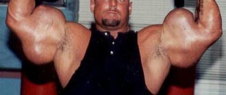
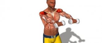
The shoulder of muscular people has the appearance of a roll, flattened on the sides. The musculature of the shoulder consists of muscles located parallel to the vertical axis of the shoulder. On the front surface of the shoulder are strong forearm flexors. The skin in this area is thin, so the outlines of the muscles are clearly visible, especially when the biceps muscle contracts, which then takes on the shape of a hemisphere. It is widely believed that the larger and more convex this hemisphere, the stronger the person.
BICEPS
The biceps, or biceps brachii muscle, consists of two heads. The long head begins from the supraglenoid tubercle, and the short one from the coracoid process of the scapula. Both heads are located along the humerus. Just below the elbow they are attached to the inside of the radius. The main function of the biceps is to flex the arm at the elbow joint, as well as participate in supination of the forearm, when the palm facing down turns up. The relief of the biceps is best seen when the forearm is flexed when it is in a supinated position.
In addition to the biceps, two more muscles are responsible for bending the arm at the elbow: the brachialis and the brachioradialis.
BRACHIAL MUSCLE
The brachialis muscle is located under the biceps. It can only be seen under the inner edge of the biceps. The outer edge is visible only at the insertion of the deltoid muscle in the area of the lower half of the humerus. The development of the brachialis muscle also affects the steep outline of the biceps muscle. The brachialis muscle starts from the lower half of the anterior surface of the humerus and attaches to the tuberosity of the ulna. Thus, the brachialis muscle raises the ulna, participating only in flexion of the forearm.
BRAIRADIAL MUSCLE
The brachioradialis muscle starts from the humerus, runs along the entire forearm and is attached to the radius in the area of the wrist joint. The main function of the brachioradialis muscle is to flex the arm at the elbow joint. When flexing the forearm, especially if this movement occurs while overcoming any resistance, the brachioradialis muscle clearly protrudes in the form of a sharp ridge in the area of the cubital fossa.
TRICEPS
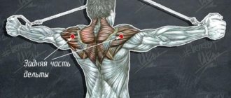
On the back surface of the shoulder, the triceps brachii muscle stands out - Triceps or triceps brachii muscle. As the name of the muscle itself indicates, it has three heads. The long head starts from the subarticular tubercle of the scapula, the medial (inner) and lateral (lateral) heads start from the humerus. All three heads converge into one tendon, which attaches to the olecranon process of the ulna.
All three heads of the triceps cover the elbow joint, and its long head also covers the shoulder joint. The main function of the triceps is to extend the arm at the elbow joint. The muscle is visible when trying to straighten the arm at the elbow joint, performed with resistance: then the outer and long heads in the upper half of the shoulder become noticeable, which form a characteristic fork.
We invite you to read: Wrist hygroma: causes, treatment without surgery and photos
Body parts in English: hands
Today we continue to study the names of body parts in English. And we came to the hands - one of the most important parts of the human body. So let's look at the English words related to hands.
Shoulder - shoulder
Let's start right at the top. Shoulder, shoulder is the part that connects the arm and torso. If in Russian someone cries “into his vest,” then in English they use the shoulder for this - substituting shoulder to cry on. But look over someone's shoulder, this is the same as in Russian “to look over your shoulder” - to, for example, peek at your phone password.
Upper arm – ?
We go down from the shoulder to the part of the arm that is between the shoulder and elbow. What is this part of the hand called in Russian? There will be a small problem with the answer. The point is that there is confusion in Russian here. From an anatomical point of view, this is the “shoulder”.
And what we call a “shoulder” in everyday life is actually a “shoulder joint”. I can hardly imagine how orthopedists in hospitals cope with such confusion, but apparently they do.
But in ordinary, everyday Russian language, it turns out for upper arm and there is no suitable word.
Armpit - armpit
This part of the body usually brings us nothing but inconvenience - at a minimum we need to buy deodorant, or even a razor. And its name is strange - armpit.
Where do mice come from? It turns out that it was named so not because of the mice, but because of the muscles. Armpit - under the muscles. But the muscles are already named because of mice, but that’s another story.
There are no mice or muscles in English naming. Armpit - that is, literally, a hole in the hand.
Elbow – elbow
The next word is elbow. This word is used in an interesting expression - elbow grease, that is, “elbow grease.”
What is this magical substance? When the English say you need to add a little elbow grease, they mean that you need to roll up your sleeves and work hard.
And elbow can also be a verb, for example to elbow your way through the crowd - to make your way through the crowd, working with your elbows.
Forearm - forearm
The next part of the arm is between the elbow and the hand, and it is called the forearm. In Russian - forearm.
Wrist - wrist
The place where the hand is attached to the rest of the arm, and also where bracelets are worn and the pulse is measured, is the wrist. Pay attention to the pronunciation of the English word wrist - the letter w is not readable, just like, for example, in the word write, to write. You may have heard about wristwatch - a watch for the wrist, but in our opinion - a wristwatch.
And now we come to hand - this word, depending on the situation, can mean both an arm and a hand. Let's see what's interesting here.
Palm - palm
It is written and read the same as “palm”, but this is just a coincidence, do not pay attention. Those who are younger will probably remember facepalm - a face covered with a palm, a classic gesture when you are overwhelmed with emotions caused by the oddities of the people around you.
Despite the vivid and accurate image, there is still no adequate translation in Russian: hand? facepalm? Let it remain facepalm.
Those who are older will probably remember Palm handhelds - in those distant times when phones were only for calls and SMS, and handheld computers were a separate class of devices.
Fingers - fingers
The significant difference between fingers and fingers is that if in Russian these are the fingers of both the hands and feet, in English there is a separate word for toes - which we will talk about in the material about feet, and fingers is exclusively that what's on hand. Just like in the Russian language, each of the fingers has its own name, which we will also talk about separately.
Fist - fist
If you put all your fingers together you get a fist. There are various interesting words with fist, but I will mention fist bump, also known as bro fist. As far as I know, there is no adequate Russian name for this gesture. And there is also a fist pump, a victory gesture when something has been achieved and you say “YES!”
Knuckles - knuckles
And the last word is knuckles. I don't know if this part of the body has an official name, but I know it as the knuckles - the bones that stick out when you make a fist.
Some cuisines have dishes that have knuckes in their names, for example pork knuckles, known to us as “knuckle”. There is also a funny expression knuckle sandwich.
When you promise someone such a treat, you mean that you will give that person a beating.
One last word before I completely finish with my arms, and before I start talking about my legs.
What word can be used to describe both arms and legs at the same time? In Russian there is a word for this, “finiteness” (apparently, everything else is “infinity”), which, although applicable to humans, somehow immediately brings to mind biology lessons. In English, “arm and leg” is a slightly more euphonious word limb.
That's probably all the English words about the main parts of the hands. Next time we will finish our conversation about vocabulary related to the human body by talking about the remaining limbs - the legs. Happily.
Source: https://english99.ru/bodyparts-3-arms/
Metacarpal bones
This section, consisting of 5 tubular metacarpal bones, also demonstrates the intricate structure of the hand. The skeleton of these tubular bones is complex. Each of them has a body, a base and a head. The metacarpal bone of the 1st finger is shorter than the others and is massive. The second metacarpal bone is the longest. The rest decrease in length as they move away from the first and approach the ulnar edge.
The bases of the aforementioned metacarpal bones articulate with the bones that form the carpus. The first and fifth metacarpal bones have bases with saddle-shaped articular surfaces, the others are flat. The heads of the metacarpal bones, having an articular surface (hemispherical), articulate with the proximal digital phalanges.
MUSCLES OF THE FOREARM
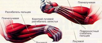
The forearm in its normal state is club-shaped with a flattened anterior and posterior surface. In the upper part of the forearm there are, for the most part, the belly of the muscles, in the lower part, mainly their tendons. In muscular people, the shape of the forearm can change to a large extent due to muscle contraction.
From an anatomical point of view, the muscles of the forearm are divided into three groups. Some of them are responsible for wrist movements, while others are responsible for finger movements. In front, on the side of the palm, there is a group of flexors. On the opposite side are the extensors. The third group of muscles is located in the area of the thumb.
- Palmaris longus muscle
- Flexor carpi radialis
- Flexor carpi ulnaris
- Flexor digitorum superficialis
- Flexor digitorum profundus
- Flexor pollicis longus.
flexor carpi radialis, palmaris longus, flexor digitorum superficialis, flexor carpi ulnaris. The pronator starts from the inner condyle of the humerus and is attached in the wrist to the phalanges of the fingers from the palmar surface of the hand. The above muscles form elongated muscle ridges, which are noticeable when the hand is bent at the wrist towards the palm and little finger.
- Extensor carpi radialis longus
- Extensor carpi radialis brevis
- Extensor carpi ulnaris
- Extensor digitorum
- Extensor pollicis longus
- Extensor pollicis brevis
- Extensor index finger
The extensors are located on the back of the forearm. Covered only by thin skin, they are clearly visible in muscular people. The prominent muscles primarily include the extensor muscles of the little finger and index finger, the extensor carpi ulnaris, the abdomen of which is especially clearly visible along the edge of the ulna.
In addition, the same group of muscles includes the long and short extensors of the thumb and its long abductor muscle. All of the above muscles make it possible to bend the hand towards its back, move the hand towards the thumb and little finger, and extend the fingers. Other muscles are grouped near the radius.
We suggest you read: Diseases of the hip joint: symptoms, treatment, diagnosis
Proportions
Now let's talk about individual finger lengths. The basic unit will be the index finger, P2. So how does it compare to other fingers:
- Maximum distance between P1 and P2 = 1.5.
- The maximum distance between P2 and P4 = 1. P3 can move away from P2 by the same distance.
- Maximum distance between P4 and P5 = 1.
- The maximum angle between P1 and P5 = 90°.
These values are approximate and may vary from person to person. Deviations from the norm, no doubt, happen, but if you need a universal scheme, feel free to use this one.
Wrist joint
It consists of the radius and the bones of the proximal row of the wrist: triquetrum, lunate and scaphoid. The ulna is complemented by an articular disc and does not reach the wrist joint. The main role in the formation of the elbow joint is played by the ulna. Whereas the wrist is radial. The wrist joint is elliptical in shape.
It allows abduction, adduction, flexion and extension of the hand. A small passive rotational movement (10-12 degrees) is also possible in this joint, but is carried out due to the elasticity of the articular cartilage. Through the soft tissues it is easy to detect the gap of the wrist joint, which can be palpated from the ulnar and radial sides.
The movements of the wrist joint are closely related to the work of the midcarpal joint, located between the distal and proximal rows. Its surface is complex and irregular in shape. When flexing and extending, the range of mobility reaches 85 degrees. The adduction of the hand in the above-mentioned joint reaches 40 degrees, abduction – 20. The wrist joint can perform circumduction, i.e. Roundabout Circulation.
This joint is strengthened by numerous ligaments. They are located between individual bones, as well as on the lateral, medial, dorsal and palmar surfaces of the wrist. The collateral ligaments (radial and ulnar) play the most important role. On the ulnar and radial sides, between the bony elevations, there is a flexor retinaculum - a special ligament.
Main muscle groups
The main muscle groups are:
- back muscles;
- chest muscles;
- shoulder muscles;
- arm muscles;
- abdominal muscles;
- leg muscles.
The musculature of the hip joint is represented by fibers of various types and functionality. This is primarily due to the varied trajectory of movement that the hip can perform. So, if we classify muscle fibers into groups according to function, in the anatomy of the hip joint we should highlight:
- The transverse, or frontal, muscle group, which is responsible for flexion and extension of the lower limb in the pelvic area. Among them are the flexor muscles (sartorius, iliopsoas, pectineus, rectus, tensor fascia lata) and hip extensor muscles (gluteus maximus, adductor magnus, semitendinosus, semimembranosus and biceps). Thanks to their coordinated work, a person can sit down and stand up, squat down and take a vertical position, pull his legs to his chest and straighten up.
- The anteroposterior, or sagittal, muscles regulate the adduction and abduction of the leg. This group includes adductors (major, short and long adductors, gracilis and pectineus) and abductors (obturator internus, tensor fascia lata, twin, piriformis, gluteus medius and minimus) muscle fibers.
- The longitudinal muscle group coordinates the rotation of the hip. Here the supinator muscles are distinguished (gemini, piriformis, iliopsoas, quadratus, sartorius, obturator, gluteus maximus and posterior groups of the middle and small gluteal fibers) and pronators (tensor fascia lata, semitendinosus, semimembranosus, anterior group of the middle and small gluteal fibers) .
Each of the muscles represented in the anatomy of the hip joint performs not only a motor function: powerful fibers take on part of the load during movements. And the more trained they are, the better they cope with pressure, thereby unloading the joint and performing a shock-absorbing function. Thanks to this, the likelihood of injury due to unsuccessful movements is also reduced, since the muscles are more mobile and extensible than the tissues of the joint.
Each individual muscle is an integral organ consisting of many small muscle fibers - myocytes, as well as dense and loose connective tissue in varying proportions. It has 2 functional zones: the abdomen and the tendon. The abdomen performs mainly a contractile function, therefore it is represented by a combination of connective tissue and myocytes capable of contraction and excitation.
Innervation and blood supply to each muscle is carried out due to the thinnest capillaries and nerve fibers located between bundles of 10–50 myocytes. Thanks to this, muscle tissue receives the necessary nutrition, is supplied with oxygen and nutrients, and can also contract in response to an impulse transmitted by nervous tissue.
Each muscle fiber looks like a long multinucleated cell, the length of which is several times greater than the cross section. The membrane covering the myocyte combines a different number of small myofibrils, depending on the number of which, white and red muscles are distinguished. In white myocytes, the number of myofibrils is higher, so they respond faster to impulses and contract more actively. Red fibers belong to the group of slow fibers because they have fewer myofibrils.
Each myofibril consists of a number of substances on which the functional characteristics and properties of muscles depend:
- Actin is an amino acid protein structure capable of contraction.
- Myosin is the main component of myofibrils, formed by polypeptide chains of amino acids.
- Actinomyosin is a complex of protein molecules actin and myosin.
The main part of myocytes consists of proteins, water and auxiliary components: salts, glycogen, etc. Moreover, the majority is water - its percentage ranges from 70–80%. Despite this, each individual muscle fiber is extremely strong and resilient, and this strength increases depending on the number of myocytes combined into the muscle.
The anatomical structure of the knee joint includes all the key elements of the musculoskeletal system: nerve fibers, muscles, ligaments and, of course, osteochondral structures. To understand how this mechanism works, you should carefully study each of these elements, its structural features and role in the mobility of the lower extremities.
The knee consists of three bones:
- Femoral. It is attached to the joint at the distal end and serves as a kind of support for the leg.
- Tibial. This tubular bone is adjacent to the knee at its proximal end and is primarily responsible for the mobility of the limb.
- Patella, or kneecap. The largest sesamoid bone in the human body protects the knee joint from possible injuries resulting from lateral displacement (for example, due to an unsuccessful dislocation, twisted leg, and other similar injuries).
By the way, a normal patella does not form in humans right away: in infancy, this bone is not yet sufficiently developed and is represented by elastic cartilaginous formations. This anatomical feature protects mobile fidgets from serious injuries: during periods of active crawling and frequent falls, elastic cartilage prevents bone damage, however, the risk of a patella fracture is significantly reduced.
From below, the anatomy of the knee is represented by cartilaginous condyles, which are in contact with the surface of the tibial plateau, contributing to the correct formation of a special recess. It is this depression that is the key link in the mechanism of flexion and extension of the knee joint.
Since the adjacent tubular bones that form the knee are disproportionate in either area or surface shape, something is needed between them that will compensate for this incompatibility, acting as a kind of shock absorber. This is precisely the role played by the menisci - small flexible formations that maintain the stability of the joint, evenly distributing the load on the adjacent surfaces of the bones. The free edges allow them to move freely in the joint cavity.
Despite the fact that the anatomical structure of the menisci resembles cartilage tissue, and in many reference books they are classified specifically as cartilage, the formations themselves are slightly different from ordinary cartilage: they are more flexible because they include a high percentage of elastin fibers. It is thanks to this that they manage to ensure full interaction of bones under high load, preventing their abrasion and deformation. Therefore, with the slightest injury to the meniscus, the entire joint, including bone structures, suffers.
Knee ligaments
The ligamentous apparatus of the knee joint serves as a strong mechanism that holds each bone in a certain position, without limiting the possible trajectory of movement. It is thanks to the ligaments that the knee does not “scatter” at the first unsuccessful step, maintaining its configuration and functionality.
Ligaments located in the area of the knee joint are represented by the following groups:
- lateral - collateral perioral and tibial;
- posterior - patella, medial and lateral suspensory, popliteal, arcuate;
- intra-articular - transverse and two cruciform.
Despite the fact that each of these groups is functional and irreplaceable in its own way, the cruciate ligaments - the anterior and posterior - are of greatest importance for joint mobility. The anterior cruciate ligament fibers support the knee joint, fix the lateral condyle of the surface of the tibia and prevent excessive forward displacement of the tibia, which, in turn, helps protect the joint from serious injury.
It is quite difficult to stretch, let alone tear, the cruciate ligaments: they are located inside the knee itself and are reliably protected by adjacent tissues. However, with inadequate physical activity and a pathological trajectory of movement, such an injury is quite possible, so you should be careful and have a reasonable approach to scheduling exercises, because knee restoration in this case is an extremely long and labor-intensive process.
Alternate contraction and relaxation of the muscles causes the knee to move in three planes, thereby ensuring mobility and stability of the lower limb. That is why the main classification of the muscular system is based not on the anatomy or localization of each group, but on the functions assigned to it:
- Knee flexion. This movement is ensured by the balanced and complete work of the largest muscle group of the knee joint. It includes the biceps, semitendinosus, semimembranosus, popliteal, gastrocnemius, plantaris, sartorius and gracilis muscles.
- Extension of the joint. This function is assigned to only one, but the largest muscle of the leg - the quadriceps. It consists of the direct, lateral, medial and intermediate wide muscle fibers.
- Pronation is the inward movement of the leg. Limited “rolling” of the lower leg towards the internal axis is ensured by the popliteal, semitendinosus, gracilis, sartorius, semimembranosus, and medial head of the gastrocnemius muscles.
- Supination is an outward movement. Turning the tibia outward is possible due to the contraction of the biceps and lateral head of the gastrocnemius muscle.
https://www.youtube.com/watch?v=upload
The nerve fibers of the knee joint are a complex interconnected network, which ensures the full functioning of the lower extremities. Despite the fact that the innervation network of the knee is not very developed, each of its elements plays a key role, which means that at the slightest failure the entire system of joint mobility is “switched off”.
The nervous system, localized in the knee area, is represented by the following fibers:
- The meniscal nerve bundles penetrate the tissue along the periphery of the body of the cartilage itself, along the blood vessels of the knee. These nerves contribute to the formation of soft and soft fibers, maintaining normal innervation of joint tissues.
- The tibial nerve, through its articular branches, provides sensation to the posterior surface of the knee.
- The peroneal nerve innervates the anterior part of the knee, including the cap.
Carpometacarpal joints
They are flat in shape and inactive. The exception is the thumb joint. The range of motion of the carpometacarpal joints is no more than 5-10 degrees. They have limited mobility because the ligaments are well developed. Located on the palmar surface, they form a stable palmar ligamentous apparatus that connects the bones of the wrist and metacarpals.
There are arcuate ligaments on the hand, as well as transverse and radial ones. The capitate bone is central to the ligamentous apparatus; a large number of ligaments are attached to it. The palmar ones are much better developed than the dorsal ones. The dorsal ligaments connect the bones of the wrist. They form thickening capsules that cover the joints located between these bones. The interosseous are located in the second row of carpal bones.
In the thumb, the carpometacarpal joint is formed by the base of the first metacarpal and polygonal bone. The articular surfaces are saddle-shaped. This joint can perform the following actions: abduction, adduction, reposition (backward movement), opposition (opposition) and circumduction (circular movement).
Structure of the hand: metacarpophalangeal joints
These joints of the hand are formed by the heads of the metacarpal bones with the participation of the bases of the proximal phalanges of the fingers. They are spherical in shape, have 3 axes of rotation perpendicular to each other, around which extension and flexion, abduction and adduction, as well as circular movements (circumduction) are carried out.
We invite you to read: Treatment of finger joint diseases with folk remedies
Adduction and abduction are possible by 45-50 degrees, and flexion and extension by 90-100. These joints have collateral ligaments located on the sides that strengthen them. Palmar, or accessory, are located on the palmar side of the capsule. Their fibers are intertwined with the fibers of the deep transverse ligament, which prevents the heads of the metacarpal bones from diverging in different directions.
But hands are different
The structure of the hands is individual, just like facial features. Men's hands are different from women's, young from old, and so on. Below are a few classifications, but they do not cover the full spectrum of hand variety. A hand can have a character - and this is the right word when talking about their differences. How would you describe the hands you saw today? Tender, rough, wet, dry?...
Hand shapes
Difference in proportions of fingers and palm:
Finger shapes
And nails are different from person to person! Even with the same size of the nail plate, you can apply the desired manicure.
Synovial and fibrous sheaths of the tendons of the fingers
The extensor retinaculum, like the flexor retinaculum, plays a huge role in strengthening the position of the tendons of the muscles running underneath them. This is especially true when working with the hand: when extending and bending it. Nature has designed a very competent structure of the hand. The tendons find support in the above-mentioned ligaments from their inner surface. Separation of tendons from bones is prevented by ties. This allows you to withstand high pressure during intense work and strong muscle contraction.
Special tendon sheaths, which are bone-fibrous or fibrous canals, help reduce friction and slip of the tendons running to the hand from the forearm. They contain synovial vaginas. The largest number of them (6-7) is located under the extensor retinaculum. The radius and ulna have grooves that correspond to the location of muscle tendons. And also the so-called fibrous bridges, which separate the canals from each other and pass to the bones from the extensor retinaculum.
Palmar synovial sheaths belong to the flexor tendons of the fingers and hands. The common synovial sheath extends to the center of the palm and reaches the distal phalanx of the fifth finger. The tendons of the superficial and deep digital flexors are located here. The thumb has a long flexor tendon, located separately in the synovial sheath and passing onto the finger along with the tendon.
Lines are the basis of authenticity
Before drawing a human hand, analyze again what parts the hand consists of. Remember that the contours of the palm and fingers, taking on specific shapes in the drawing, become more and more rounded. For example, the line connecting the fingers and palm is shaped like an arc, as is the outline of the hand itself—different lengths of the fingers create a semicircle when drawing fingers pressed together. The thumb is slightly turned in relation to the rest of the palm; its contour will not be straight, but somewhat rounded.
Muscles of the small finger
This group of muscles forms an elevation on the inside of the palm. These include: the abductor digiti minimi, the opponens minimi, the palmaris brevis, and the flexor brevis muscle.
They originate from nearby bones in the wrist. These muscles are attached to the base of the fifth finger, more precisely its proximal phalanx, and to the fifth metacarpal bone. Their function is reflected in the name.
In the article we tried to most accurately represent the structure of the hand. Anatomy is a fundamental science that, of course, requires more careful study. Therefore, some questions remained unanswered. The structure of the hand and wrist is a topic that interests not only doctors. Knowledge of it is also necessary for athletes, fitness instructors, students and other categories of people. The structure of the hand, as you noticed, is quite complex, and you can study it for quite a long time, relying on various sources.
What does it look like?
It will be easier to draw if, while sketching a sketch, you decide what the subject of the image looks like - something simple, even primitive. Do you agree that the human hand is similar to a shovel not only in appearance, but also in functionality? You can start a sketch with this - draw a contour similar to a shovel: the wrist is the handle of the shovel, and the contour of the palm with fingers is its canvas. It’s difficult to immediately decide how to draw a hand with a pencil step by step, which is why it’s worth starting with a basic sketch.