For students of sports universities , athletes and all the curious, we explain in simple language the sources of energy and the physiology (mechanism) of muscle contraction, as well as the structure of muscles, with simple formulas and photos, the understanding of which will help you train in the gym in the future.
600 muscles in the human body , and each of them is responsible for one or another type of activity, for example, the back ensure an even posture, keep the body in an upright position, and thanks to the extraocular muscles, we can direct our gaze in the direction in question.
With the help of muscles, we understand the world around us, the surrounding reality , if not for them, we would never have learned to walk , write, speak, or express our emotions . However, in order for muscles to function normally, they must be supplied with energy, useful nutrients, and in short, the digestion process is as follows:
After mechanical grinding in the mouth, food passes through the pharynx into the esophagus , then into the stomach , where under the influence of gastric juice (mainly hydrochloric acid and the enzyme pepsin), proteins and partly fats are broken down and enters the pylorus , then the food mass passes into the duodenum ( the beginning of the small intestine), where, under the influence of pancreatic juice and bile, further decomposition of proteins, carbohydrates and fats occurs, then the chyme passes into the small intestine, in which the final decomposition of amino acids, fatty acids and monosaccharides occurs and, due to the presence of villi, are absorbed into the walls of the small intestine , everything that is not absorbed passes into the large intestine, in which the processes of crushing fiber and chyme begin, as well as the absorption of residual water and the formation of feces.
Absorbing nutrients through the stomach and intestines, the resulting substances enter the blood (portal vein ), and then go to the liver , after which they enter the general bloodstream , washing all human tissues.
Human muscular system
The human muscular system allows you to coordinate the movements of the body, keep it in balance, breathe, as well as transport food and blood within the body ; in addition, it protects the insides from damage, and also acts as a converter of chemical energy into mechanical and thermal.
There are only three types of muscles in the human body:
- skeletal
- smooth
- heart muscle
Human muscular system (A - cardiac muscle, B - skeletal muscle, C - smooth muscle)
Skeletal muscles
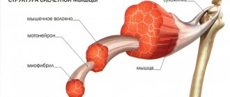
Human skeletal muscle, also known as striated muscle , is attached to the bones and consists of fibers, which in turn consist of muscle cells. Each muscle cell has two nuclei , which are responsible for division and repair. so-called myofibrils (threads), which are contained in muscle cells . The number of myofibrils in a muscle cell can reach up to several thousand. Thus, muscle cells form tissue , which in turn forms muscle.
Our skeletal muscles contain fibers, nerve endings and blood vessels. Muscle contraction occurs with the help of nerve impulses that come from the spinal cord to muscle tissue, that is, the transmission of the nerve impulse occurs along the path - brain → spinal cord → the muscles we need. Now we understand why spinal cord injury is so dangerous.
A person regulates the intensity of muscle contraction using the strength of the impulse supplied along the nerve endings.
Human skeletal muscles
Smooth muscle
Smooth muscle performs involuntary contractions, consists of spindle cells, being one of the most important components of the muscular hollow organs, as well as an integral part of the blood and lymphatic vessels, helps transport the contents of the hollow organs (transport food to the intestines), constricts the pupil, adjusts blood pressure, and other processes that occur involuntarily.
All smooth muscle contractions do not cause fatigue and are regulated by the autonomic system (autonomic nervous system, which is responsible for the functioning of internal organs).
You can train smooth muscles, for example, by increasing endurance , you improve the functioning of the cardiovascular system.
Smooth muscle
Heart muscle
The heart continuously contracts throughout life, providing movement, pumping blood, nutrients, and other vital substances through the vessels to the tissues of the body. Acting as a pump , the heart operates in a mode of continuous, rhythmic, single contractions.
The structure of myocardial resembles the structure of skeletal muscles, which also contain myofibrils consisting of actin and myosin, including the troponin-tropomyosin protein complex.
Picture of the heart, which shows the structure of the heart where you can see the myocardium
The mechanism of muscle contraction of the heart occurs for the same reasons as in striated muscles, thanks to Ca2+ (calcium) ions that are released from the sarcoplasmic reticulum (membrane organelle of muscle cells), only in this case it is less ordered (compared to skeletal muscles ).
Heart muscle and its structure
Mechanical and metabolic load
It is well known that physical adaptation to exercise, including muscle growth, results from the application of acute program variables. There is no doubt that resistance training leads to muscle growth, however, scientists are still not sure what exactly causes muscle growth. Resistance training produces two specific types of stress, mechanical and metabolic, and both can provide the necessary stimulus for muscle growth (Bubbico and Kravitz, 2011). Brad Schoenfeld is a scientist and the author of two comprehensive reviews on training for muscle growth. “Mechanical tension is by far the primary driver of muscle growth from exercise,” Schoenfeld explains. “There is compelling evidence that metabolic stress also promotes adaptive hypertrophy. The challenge for research is that mechanical and metabolic stress act in tandem, making it difficult to isolate the influence of each” (Schoenfeld, 2013).
Mechanical stress is the stress from physical activity applied to the structures of the motor neuron and the fibers attached to it, together usually called motor units. Resistance training causes microtrauma to muscle tissue, which sends signals to satellite cells responsible for repairing mechanical structures after damage, as well as for the formation of new muscle proteins (Schoenfeld, 2013; 2010).
Additionally, in his study on cellular adaptation to resistance training, Spangenburg (2009) confirms that “mechanisms activated by exercise result in changes in muscle signaling pathways that are responsible for hypertrophy.”
Metabolic stress occurs as a result of the muscle producing and consuming the energy needed to support contractions. Moderate-intensity, high-volume training programs that lead to muscle growth use the glycolytic system to produce energy. By-products of anaerobic glycolysis: the accumulation of lactate and hydrogen ions - lead to changes in blood acidity and cause acidosis. Research shows a strong connection between blood acidosis and increased levels of growth hormones that support muscle protein synthesis. In a review of research by Bubbico and Kravitz (2011), Fr.
When developing a training program that aims to increase muscle mass, you need to know how to use the stress of exercise without creating a negative combination with other stressors. A good personal trainer should know how to adjust exercise loads to promote optimal results from the training program. It is necessary to design a resistance training program using the correct variables of exercise intensity, repetition range, and rest intervals to create mechanical and metabolic loads on muscle tissue that stimulate the production of hormones and promote the synthesis of contractile proteins responsible for muscle growth (Schoenfeld, 2013; Bubbico and Kravitz, 2011).
https://youtu.be/BCGAj_QLPOg
The structure of human muscles (muscle fibers)
Human muscles are made up of muscle fibers , which in turn are made up of muscle cells. Taken individually, a muscle fiber is a multinucleated muscle cell, the diameter of which varies from 10 to 100 microns a sarcolemma (cell membrane) shell sarcoplasm (the contents of the cell, the basis of which is the matrix). Myofibrils are located in sarcoplasm, that is, sarcoplasm fills the space between myofibrils and surrounds the cell nuclei. The myofibril is a filament-shaped formation consisting of sarcomeres (the contractile apparatus of the muscle).
Structure of skeletal muscle
Depending on the number of myofibrils, white and red muscle fibers are distinguished.
White muscle fibers differ from red ones by having a larger number of myofibrils and less sarcoplasm; this ratio ensures the rapid contraction of white fibers. Due to the presence of myoglobin (oxygen-binding protein) in muscles, which gives color, muscle fibers are called red .
Sarcoplasm in muscle cells contains, in addition to myofibrils, also mitochondria (energy stations of cells in which ATP is synthesized), ribosomes, Golgi complex, fatty inclusions, and other permanent components of the cell, without which its existence is not possible ( organelles ).
The transmission of impulses within muscles occurs due to the sarcoplasmic reticulum . The basic contractile unit of striated muscles (sarcomeres) contains thick myosin filaments, formed by the protein myosin, and thin actin filaments, formed by the protein actin.
Actin , a contractile protein that accounts for about 15% of all muscle protein, is found in the thin filaments of skeletal muscles, ensuring the motor functions of cells.
Myosin is the main protein that makes up muscle fibers, thanks to which muscles have elasticity and are able to contract. The mass of myosin makes up about 55% of all contractile proteins contained in muscle fibers.
Myosin is concentrated in striated muscles (skeletal muscles), which are responsible for reflexes and purposeful movements. Thanks to the ability of myosin to break down ATP, the chemical energy of the macroergic bonds of ATP is converted into the mechanical energy of muscle contraction.
The structure of human muscles
Actomyosin , a complex consisting of the proteins actin and myosin, creates muscle fibers that are distributed in a specific order. Contraction of actomyosin is possible thanks to the energy that is released as a result of the interaction of ATP with water ( hydrolysis ), thus actomyosin determines the ability of muscles to contract (muscle contraction).
Weight training
Resistance training involves exercising additional weights to improve the appearance and functionality of skeletal muscles. Such training can increase muscle size and strength at the same time. But at the same time, there are clear differences between training that promotes muscle growth and training aimed at developing maximum effort.
Weight training by itself does not lead to muscle growth, but the training load received in the process causes fatigue, which stimulates the physiological mechanisms responsible for the growth of muscle mass. When building a program for such training, you need to take into account that the physical impact received on them must be of very high intensity, incomparable to that which the body usually receives.
As a result of weight training, the volume of muscle fibers increases, which leads to a gain of muscle mass, and the volume of fluid contained in the sarcoplasm of muscle cells also increases. What does understanding the process of adaptation of the muscular system to resistance training provide? First of all, it helps in choosing the best training method to build muscle more effectively.
The research available today explains the mechanism of the body's response to stimuli affecting it. However, each person may experience different results in response to the same effects of resistance exercise.
The ability to increase muscle mass and lean muscle mass depends on many variables: age, gender, experience with similar training, genetics, sleep and nutrition patterns, and the amount of fluid consumed. Physical and emotional stress also affect the adaptation to training of physiological systems and, as a result, weight gain. Thus, insufficient sleep and work overload can have a negative impact on muscle growth.
Knowledge about this science can help you achieve maximum results.
Mechanism of muscle contraction
Muscle contraction occurs due to the sliding of actin filaments along the myosin filaments (sliding contraction), while the total length of the actomyosin complex , without changing the length of the actin and myosin filaments. A primary role in muscle contraction is played by temporarily closed cross bridges (heads of myosin molecules), which ensure the movement of actin filaments along the myosin filaments. Accordingly, the force of muscle contraction will depend on the bridges - the more bridges are attached to the actin filaments, the stronger the contraction.
The energy required for this “ sliding contraction ” is provided by the interaction of ATP with actomyosin, resulting in adenosine triphosphate (ATP) breaking down into adenosine diphosphate (ADP) and phosphoric acid (H3PO4).
Along with ATP, calcium, magnesium and water also play an important role in muscle fiber contraction .
The amount of water that muscle is approximately 72-80% , while muscle tissue also contains a large amount of proteins , and in small quantities glycogen , phospholipids, cholesterol, creatine phosphate, creatine, and vitamins.
The more muscle fibers there are in skeletal muscles, the stronger they will be.
The mechanism of human muscle contraction
Metabolic and energy processes of skeletal muscles
Thanks to creatine phosphate, the body has the energy to restore ATP reserves.
During the rest period, muscles contain approximately 5 times more creatine phosphate compared to ATP. After the start of muscle contraction, the number of ATP molecules rapidly decreases, and ADP increases. The reaction to produce ATP from creatine phosphate occurs quite quickly, but the number of ATP molecules that can be synthesized directly depends on the initial level of creatine phosphate. Muscle tissue also contains a substance called myokinase. Under its influence, two ADP molecules are converted into one ATP and ADP. The reserves of ATP and creatine phosphate are in total sufficient to work the muscles at maximum load for 8 to 10 seconds.
Energy of muscle contractions
As we already know, muscle contraction is the movement ( opening-closing ) of transverse bridges, and naturally, this process requires energy supply. However, reserve in the muscles is very small, so it must be constantly “drawn” from somewhere and restored.
The main and main supplier of energy, thanks to which the body is supplied with energy, is the ATP molecule, which is broken down thanks to the heads of myosin bridges, thus generating energy for muscle contraction. However, a muscle cell contains a limited supply of ATP molecules; their quantity is enough for only 8 repetitions , the first 2 seconds:
- ATP + H2O = ADP + H3PO4 + energy
The problem of limited energy supply for muscle contraction is easily solved by resynthesis or re-creation of ATP molecules.
ATP - as a universal source of human energy
Sources of muscle energy
Depending on the time the muscles are under load , as well as on the degree of intensity of performing a particular exercise, the following types of systems that provide energy to the muscles:
- Phosphogenic (ATP and KrP)
- Glycolytic (anaerobic and aerobic glycolysis)
- Oxidation (carbohydrates and lipids are broken down with active oxygen supply)
In some cases, with a long-term absence of consumption of carbohydrates and fats amino acids can act as an energy source , thanks to the process of gluconeogenesis , in which amino acids are converted into pyruvate , the total formula is below:
2 Pyruvate + 4ATP + 2GTP + 2NADH + 2H+ + 4H2O → glucose + 4ADP + 2GDP + 6Pi + 2NAD+
Each energy system is characterized by power and capacity.
Power is an indicator that limits the intensity of physical work performed and characterizes the maximum amount of energy that is released per unit of time, as well as the maximum amount of resynthesized ATP per unit of time.
In turn, the capacity of energy systems limits the duration of muscle work.
Sources of muscle energy
Creatine phosphate breakdown
CrP molecule reduces ADP (adenesine diphosphate) to ATP (adenosine triphosphate), by transferring the phosphate group from CrP to ADP to form creatine and ATP:
- ADP + KrP ⇆ ATP + creatine
Thus, creatine phosphate is responsible for a constant level of ATP concentration, due to a reversible rephosphorylation .
Basically all creatine phosphate is found in skeletal muscles , heart, brain and nerve tissues.
The formation of free creatine stimulates anaerobic glycolysis in slow muscle fibers ( SMF ), and aerobic glycolysis in fast muscle fibers ( FMF ).
For bodybuilding , an important role will be played by the fact that complete restoration of creatine phosphate reserves occurs within 3-5 minutes , and this is possible only after the cessation of intense strength training, that is, in the intervals between approaches. For example, if Creatine Phosphate levels were restored during a strength exercise, we could perform the set for a very, very long time, but this does not happen, due to the limited amount of creatine phosphate in the muscles and its recovery method.
In the absence of motor activity, that is, when the muscles are in a calm state, the concentration of CrP is 3-8 times higher than the concentration of ATP, due to which, during short and intense periods of muscle activity , creatine phosphate successfully copes with the lack of ATP to perform muscle contractions.
Strength training performed in the gym consumes creatine phosphate 5-10 seconds pathway for ATP recovery, anaerobic glycolysis, is activated.
Explosive power and creatine consumption in muscles
Training the phosphate system is a top priority for all athletes who use explosive , ranging from fast jogging over short distances to performing a strength approach in the gym for 2-4 repetitions. Such athletes primarily include: sprinters, football players, double-athletes, wrestlers, boxers, discus/shot/javelin throwers, high/long jumpers, boxers , tennis players, in other words, all athletes who at one time or another need to show high physical performance in a short period of time.
The phosphagen system is considered a very fast, powerful source of energy replenishment (per unit time, the power of KrP is 3 times higher than the power of glycolysis, and 4-10 times higher than oxidation!), however, the capacity is not large, which is why with very intense physical work, the athlete can continue his efforts no more than 5-6 seconds .
When creatine phosphate reserves run out, glycolysis processes are started the sarcoplasm .
Anaerobic glycolysis
At about 20-30 seconds of performing a strength exercise, creatine phosphate reserves are exhausted, and to supply the body with energy, anaerobic glycolysis , that is, carbohydrates (glucose) are broken down in the absence of oxygen, to lactate (lactic acid):
- C6H1206 (glucose) + 2 H3P04 + 2 ADP = 2 C3H6O3 (lactate) + 2 ATP (energy) + 2 H2O
Due to the fact that in anaerobic glycolysis, lactic acid , which is very slowly washed out, over time it accumulates more and more in the muscles during their contraction, as a result, our further muscle movements become difficult , it is felt burning sensation , and eventually the contractions stop.
Lactic acid acts as a blocker , which disrupts the signal from motor neurons to muscles.
During intense exercise, lactate is often used by the body as a source of energy and raw material for the production of glucose .
Muscle glycogen consumption in the gym
Lactic acid is neutralized in the muscles almost completely (95%) after 1 hour 15 minutes . In order to speed up this process, we recommend using active rest and cool-down after training, since 75% of the lactic acid formed during intense exercise passes from fast muscle fibers to slow ones, which are used by them as a source of energy, and as we know, Active rest is the work of slow muscle fibers, which is why passive rest is inferior to active rest.
Aerobic glycolysis
At 80-90 seconds of muscle work, the processes of aerobic glycolysis in the mitochondria are launched, the process of which requires oxygen.
At the intermediate stage of aerobic glycolysis, two molecules of pyruvate , penetrating into the mitochondria, they are oxidized through the Krebs cycle , to carbon dioxide (CO2) and water (H2O), with the formation of 38 molecules of ATP.
- C6H12O6 + 6O2 + 38ADP + 38H3PO4 = 6CO2 + 44H2O + 38ATP
Thus, aerobic oxidation of glucose is much more effective than anaerobic oxidation, however, anaerobic glycolysis is the only way to supply the body with energy under conditions of oxygen starvation . Due to the high efficiency of aerobic glycolysis, in relation to anaerobic ( 19 times !), in tissues, when oxygen enters, the anaerobic pathway of energy supply is blocked ( Pasteur ).
Types of typical aerobic exercises:
- Swimming
- Middle and long distance running
- Jump rope
- Fast walk
- Skis
- Bike
Remember that at the beginning of the exercise, energy supply occurs due to anaerobic glycolysis, but after 1-2 minutes , while maintaining active movements, the energy supply already occurs due to the processes of aerobic glucose oxidation. Of course, if the work is of an explosive short nature, then for the first 5-10 seconds the energy supply occurs due to the breakdown of KrF , and then, as the intensity decreases, anaerobic glycolysis sequentially occurs, and after 1-2 minutes aerobic glycolysis.
In untrained athletes, a lot of lactic acid is formed during physical activity, which indicates a weak aerobic system , while trained athletes can boast of aerobic abilities, that is, a high ability to absorb oxygen .
Running and aerobic glycolysis
For example, unprepared athletes begin to feel quickly enough when running/biking/skiing soreness in the muscles, in the form of a burning sensation, lactic acid is formed very quickly in them, with a lack of oxygen, thus, an imbalance in the acid-base balance in the muscles occurs ( acidosis ), that is, their acidification.
With moderate, prolonged physical activity, the body is supplied with energy through the use of muscle glycogen ; over the next 30 minutes fatty acids as energy .
The more intense and shorter the duration of the load , the greater the contribution of the anaerobic process of ATP formation, and vice versa, if the load is long, of medium intensity, the more predominant are the processes of aerobic ATP synthesis.
Aerobic oxidation of carbohydrates and fats
When the body's tissues are sufficiently supplied with oxygen carbohydrates and lipids come to the rescue in energy supply are oxidized in the mitochondria to release energy.
palmitic acid as an example , the equation would look like this:
- CH3(CH2)14COOH + 23 O2 +129 ADP = 6CO2 + 146 H2O + 129 ATP
Using glucose (carbohydrate) as an example
- C6H12O6 + O2 = CO2 + H2O + Q
The capacity of the oxidative system is the largest and is many times greater than all others, for example, 20 times higher than the capacity of glycolysis, which is why, during monotonous, prolonged work, for 1-2 hours , it is the oxidative energy system that does the work.
To provide the oxidation energy system, carbohydrates and fats (lipids) are used, and the more intensely a person trains, the more carbohydrates are consumed and the less fats.
Marathon swimming is the best way to burn fat
MOC (maximum oxygen consumption)
Conventionally, in order to characterize the degree of intensity of the work performed, the term MOC (maximum oxygen consumption) was introduced, which is unique for everyone. So, for example, when performing light aerobic work (light running), fats , in this case the MIC does not exceed 50% , when the MIC reaches 70% carbohydrates begin to be consumed , that is, when performing more difficult, intense aerobic work.
MOC (maximum oxygen consumption)
Communication between the IPC and ANNO
With an increase in power anaerobic-aerobic comes into play, there is a decrease in oxygen demand and a gradual increase in lactic acid PANO 1 , if you continue to perform intensive work, the energy supply will switch to the anaerobic path of supplying energy to the muscles PANO 2 , and the higher the threshold of PANO, which is determined as a percentage of MOC, the higher the aerobic capabilities of the athlete.
If you find an error, please select a piece of text and press Ctrl+Enter.
Creatine phosphate formation process
The amount of ATP in muscle tissue is very limited and for this reason the body must constantly replenish its reserves.
This process occurs with the participation of creatine phosphate. This substance has the ability to detach a phosphate group from its molecule, attaching it to ADP. As a result of this reaction, creatine and an ATP molecule are formed. This process is called the “Lohmann reaction”. This is the main reason why athletes need to take supplements containing creatine. Please note that creatine is used only during anaerobic exercise. This fact is due to the fact that creatine phosphate can only work intensively for two minutes, after which the body receives energy from other sources.
Thus, the use of creatine is justified only in strength sports. For example, there is little point in using creatine for track and field athletes, since it cannot improve athletic performance in this sport. The reserve of creatine phosphate is also not very large and the body uses the substance only in the initial phase of training. After this, other energy sources are connected - anaerobic and then aerobic glycolysis. During rest, the Lohmann reaction proceeds in the opposite direction and the supply of creatine phosphate is restored within a few minutes.


